. 2009 Sep;23(9):1346-59.
doi: 10.1210/me.2009-0093. Epub 2009 May 21.

Apurinic/apyrimidinic endonuclease 1 alters estrogen receptor activity and estrogen-responsive gene expression

Affiliations

Apurinic/apyrimidinic endonuclease 1 alters estrogen receptor activity and estrogen-responsive gene expression

Carol D Curtis et al. Mol Endocrinol. 2009 Sep.

Abstract

Apurinic/apyrimidinic endonuclease 1 or redox factor-1 (Ape1/Ref-1) is a pleiotropic cellular protein involved in DNA repair and, through its redox activity, enhances the binding of a select group of transcription factors to their cognate recognition sequences in DNA. Thus, we were intrigued when we identified Ape1/Ref-1 and a number of DNA repair and oxidative stress proteins in a complex associated with the DNA-bound estrogen receptor alpha (ERalpha). Because Ape1/Ref-1 interacts with a number of transcription factors and influences their activity, we determined whether it might also influence ERalpha activity. We found that endogenously expressed Ape1/Ref-1 and ERalpha from MCF-7 human breast cancer cells interact and that Ape1/Ref-1 enhances the interaction of ERalpha with estrogen-response elements (EREs) in DNA. More importantly, Ape1/Ref-1 alters expression of the endogenous, estrogen-responsive progesterone receptor and pS2 genes in MCF-7 cells and associates with ERE-containing regions of these genes in native chromatin. Interestingly, knocking down Ape1/Ref-1 expression or inhibiting its redox activity with the small molecule inhibitor E3330 enhances estrogen responsiveness of the progesterone receptor and pS2 genes but does not alter the expression of the constitutively active 36B4 gene. Additionally, the reduced form of Ape1/Ref-1 increases and E3330 limits ERalpha-ERE complex formation in vitro and in native chromatin. Our studies demonstrate that Ape1/Ref-1 mediates its gene-specific effects, in part, by associating with endogenous, estrogen-responsive genes and that the redox activity of Ape1/Ref-1 is instrumental in altering estrogen-responsive gene expression.


Figures

Figure 1
Figure 1
Expression and Interaction of ERα and Ape1/Ref-1. A, Nuclear extracts (20 μg) from MCF-7, U2OS, HeLa, or MDA-MB-231 cells were separated on a denaturing acrylamide gel and transferred to nitrocellulose. Proteins were detected with Ape1/Ref-1-, ERα-, and GAPDH-specific antibodies. GAPDH was used as a loading control. B, MCF-7 cells were treated with ethanol or 10 nm E2, cell lysates were prepared, and proteins were immunoprecipitated with an antibody directed against Ape1/Ref-1. The immunoprecipitated proteins were subjected to Western blot analysis with ERα- and Ape1/Ref-1-specific antibodies. Lanes 1 and 2 contain 1% input. IP, Immunoprecipitation.
Figure 2
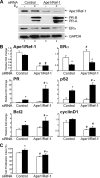
Figure 2
Effect of knocking down Ape1/Ref-1 on endogenous estrogen-responsive gene expression. MCF-7 cells were transfected with control or Ape1/Ref-1-specific siRNA and then treated with ethanol (−E2 and white bars) or 10 nm E2 (+E2 and black bars) for 24 h. A, Protein samples were subjected to Western blot analysis with an antibody that recognizes Ape1/Ref-1, PR-A and PR-B, ERα, or GAPDH. B, RNA was isolated, cDNA was synthesized, and real-time PCR was carried out with gene-specific primers. The relative fold change was determined using the comparative Ct method with the housekeeping gene, 36B4, serving as the internal control. Data from four independent experiments, which had been carried out in duplicate, were combined and are presented as the mean ± sem. C, MTT assays were performed, and absorbance measurements were collected. Cell proliferation index (%) was determined by comparing the mean absorbance of Ape1/Ref-1 siRNA samples with control siRNA. Data from three independent experiments, which were carried out in duplicate, were combined and are presented as the mean ± sem. ANOVA with a post hoc Student’s t test was used to detect significant differences in mRNA levels and cell proliferation in response to E2 (*, P < 0.05) or Ape1/Ref-1-specific siRNA (#, P < 0.05).
Figure 3
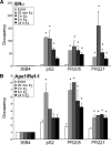
Figure 3
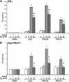
Ape1/Ref-1 association with endogenous estrogen-responsive genes. MCF-7 cells were treated with ethanol or 10 nm E2 for 20 min, 2 h, 6 h, or 24 h. Chromatin was prepared and immunoprecipitated with an antibody directed against ERα (A) or Ape1/Ref-1 (B). DNA was isolated and amplified by real-time quantitative PCR to determine whether ERα and Ape1/Ref-1 were associated with the ERE-containing region of the pS2 gene or regions of the PR gene located 205 kb (PR205) or 221 kb (PR221) upstream of the PR-B transcription start site, which contain two and one imperfect EREs, respectively. Four independent experiments, which were each carried out in duplicate, were combined and are presented as the mean number of copies of each estrogen-responsive region pulled down relative to the number of copies of the 36B4 gene region pulled down (Occupancy) ± sem. ANOVA with a post hoc Student’s t test was used to detect statistical increases in the association of ERα and Ape1/Ref-1 with these gene regions in the presence of E2 (*, P < 0.05).
Figure 4
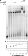
Figure 4
Effect of Ape1/Ref-1 on the ERα-ERE interaction. A, 32P-labeled oligos containing a consensus ERE were run alone (lane 1) or combined with 30 fmol baculovirus-expressed, purified ERα (lanes 2–9) and increasing (lanes 3–7) or constant (lanes 7–10) amounts of bacterially expressed, purified Ape1/Ref-1. Total protein concentrations in each lane were held constant by the addition of BSA. ERα- (lane 8) and Ape1/Ref-1- (lane 9) specific antibodies were added as indicated. Lane 10 contains purified Ape1/Ref-1 and radiolabeled ERE oligos in the absence of ERα. B, The percent of probe bound was determined from four independent experiments and is shown graphically as the mean ± sem. Statistical differences were determined using a one-way ANOVA with a post hoc Student’s t test (*, P < 0.05). Ab, Antibody.
Figure 5
Figure 5
Effect of Ape1/Ref-1 inhibitors on estrogen-responsive gene expression. MCF-7 cells were incubated with DMSO, MX, or E3330 for 1 h and then treated with ethanol (−E2 or white bars) or 10 nm E2 (+E2 or black bars) for 24 h. A, Cell lysates were prepared and separated on a denaturing acrylamide gel. The blot was subjected to Western analysis with an antibody that recognizes Ape1/Ref-1 or GAPDH. B, RNA was isolated, cDNA was synthesized, and real-time PCR was carried out with gene-specific primers. The relative fold change was determined using the comparative Ct method with the housekeeping gene, 36B4, serving as the internal control. Data from four independent experiments, which had been done in duplicate, were combined and are presented as the mean ± sem. ANOVA with a post hoc Student’s t test was used to detect significant differences in mRNA levels in response to E2 (*, P < 0.05) or the inhibitor E3330 (#, P < 0.05).
Figure 6
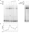
Figure 6
Effect of reduced Ape1/Ref-1 on ERα-ERE complex formation. A, 32P-labeled ERE-containing oligos were run alone (lane 1) or combined with 30 fmol baculovirus-expressed, purified ERα (lanes 2–8) and bacterially expressed, purified Ape1/Ref-1 (lanes 4–8). DTT (0.02 mm; lanes 3–7) or increasing concentrations of the Ape1/Ref-1-specific redox inhibitor E3330 (lanes 5–7) were included as indicated. Total protein concentrations were held constant by addition of BSA. B, The percent of probe bound was determined from four independent experiments and is shown graphically as the mean ± sem. C, 32P-labeled ERE-containing oligos were combined with 30 fmol ERα (lanes 1–3), 0.02 mm DTT (lanes 2–3) and wild-type Ape1/Ref-1 (lane 2) or redox-deficient Ape1/Ref-1 C65S (lane 3). Total protein concentrations were held constant by addition of BSA. Wt, Wild type.
Figure 7
Figure 7
Effect of Ape1/Ref-1 redox inhibitor on ERα association with endogenous estrogen-responsive genes. MCF-7 cells were exposed to DMSO or the Ape1/Ref-1 redox inhibitor E3330 for 1 h and then treated with ethanol or 10 nm E2 for 20 min. Chromatin was prepared and immunoprecipitated with an antibody directed against ERα (A) or Ape1/Ref-1 (B). DNA was isolated and amplified by real-time quantitative PCR to determine whether ERα and Ape1/Ref-1 were associated with the ERE-containing region of the pS2 gene or two upstream regulatory regions of the PR gene (PR205 or PR221). Three independent experiments, which were each carried out in duplicate, were combined and are presented as the mean number of copies of each estrogen-responsive region pulled down relative to the number of copies of the 36B4 gene region pulled down (occupancy) ± sem. ANOVA with a post hoc Student’s t test was used to detect statistical differences in the association of ERα and Ape1/Ref-1 with these gene regions in the presence of E2 (*, P < 0.05) or the inhibitor E3330 (#, P < 0.05).
Figure 8
Figure 8
Expression of Ape1/Ref-1 and ERα in normal human breast tissue. Normal breast tissue was subjected to immunohistochemistry using Ape1/Ref-1- (A and C) or ERα-specific (B and D) antibody. A representative slide is shown at ×10 (A and B) and ×40 (C and D) magnification. Control slides, which were not exposed to Ape1/Ref-1 or ERα antibody, are shown in the insets in A and B. Stromal (S) and epithelial (E) cells are indicated. Scale bars, 100 μm.
Figure 9
Figure 9
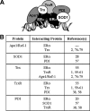
An interconnected network of oxidative stress proteins interacts with the DNA bound-ERα (A and B). Ape1/Ref-1, Trx, TrxR, PDI, and SOD1 form an interconnected network of oxidative stress proteins associated with the DNA-bound ERα.

Similar articles

Cited by

References

    1. Webster KA, Prentice H, Bishopric NH 2001 Oxidation of zinc finger transcription factors: physiological consequences. Antioxid Redox Signal 3:535–548 - PubMed
    1. Hirota K, Matsui M, Iwata S, Nishiyama A, Mori K, Yodoi J 1997 AP-1 transcriptional activity is regulated by a direct association between thioredoxin and Ref-1. Proc Natl Acad Sci USA 94:3633–3638 - PMC - PubMed
    1. Jayaraman L, Murthy KG, Zhu C, Curran T, Xanthoudakis S, Prives C 1997 Identification of redox/repair protein Ref-1 as a potent activator of p53. Genes Dev 11:558–570 - PubMed
    1. Evans AR, Limp-Foster M, Kelley MR 2000 Going APE over ref-1. Mutat Res 461:83–108 - PubMed
    1. Sweasy JB, Lang T, DiMaio D 2006 Is base excision repair a tumor suppressor mechanism? Cell Cycle 5:250–259 - PubMed

Publication types

  NODES
Association 8
INTERN 2
twitter 2