. 2009 Jul;17(1):98-109.
doi: 10.1016/j.devcel.2009.06.014.

Atg32 is a mitochondrial protein that confers selectivity during mitophagy

Affiliations

Atg32 is a mitochondrial protein that confers selectivity during mitophagy

Tomotake Kanki et al. Dev Cell. 2009 Jul.

Abstract

Mitochondrial quality control is important in maintaining proper cellular homeostasis. Although selective mitochondrial degradation by autophagy (mitophagy) is suggested to have an important role in quality control, and though there is evidence for a direct relation between mitophagy and neurodegenerative diseases, the molecular mechanism of mitophagy is poorly understood. Using a screen for mitophagy-deficient mutants, we found that YIL146C/ECM37 is essential for mitophagy. This gene is not required for other types of selective autophagy or for nonspecific macroautophagy. We designated this autophagy-related (ATG) gene as ATG32. The Atg32 protein localizes on mitochondria. Following the induction of mitophagy, Atg32 binds Atg11, an adaptor protein for selective types of autophagy, and is then recruited to and imported into the vacuole along with mitochondria. Therefore, Atg32 confers selectivity for mitochondrial sequestration as a cargo and is necessary for recruitment of this organelle by the autophagy machinery for mitophagy.


Figures

Figure 1
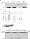
Figure 1. Atg32 is required for mitophagy
(A) Wild-type (WT) and atg32Δ strains expressing Om45-GFP were cultured in YPL medium for three days. The localization of GFP was visualized by fluorescence microscopy. DIC, differential interference contrast. (B) Wild-type (WT), atg32Δ, atg1Δ and atg11Δ strains, or (C) the atg32Δ strain transformed with a vector expressing ATG32 (ATG32(416)) or the empty vector, and co-expressing Om45-GFP were cultured in YPL or SML medium to mid-log growth phase, then shifted to SD-N medium for 0, 4 and 6 h. GFP processing was monitored by immunoblotting with anti-YFP antibody. The asterisk indicates a non-specific band. The positions of molecular mass markers are indicated.
Figure 2
Figure 2. Atg32 is a mitophagy-specific gene
(A) Wild-type (WT), atg32Δ, atg1Δ and atg11Δ strains expressing GFP-Atg8 were cultured in SMD medium to mid-log phase, and then starved in SD-N for up to 3 h. Cells were collected at the indicated time points and cell lysates were subjected to immunoblot analysis with anti-YFP antibody. (B) The wild-type (TN124), atg32Δ (TKYM131), and atg1Δ (HAY572) strains were grown in YPD and shifted to SD-N for 1.5 and 3 h. Samples were collected and protein extracts assayed for Pho8Δ60 activity. The results represent the mean and SD of three experiments. (C) Wild-type, atg32Δ, atg1Δ and atg11Δ strains were cultured in YPD medium and analyzed for prApe1 maturation by immunoblotting to monitor the Cvt pathway during vegetative growth. The positions of precursor and mature Ape1 are indicated. (D) Wild-type, atg32Δ, and atg1Δ strains were integrated with GFP at the PEX14 locus. Cells were grown in oleic acid-containing medium for 19 h, then shifted to SD-N for the indicated times. Samples were collected and analyzed by immunoblot with antibodies to YFP.
Figure 3
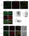
Figure 3. Atg32 localizes to mitochondria and is imported into the vacuole along with mitochondria during starvation
(A) A strain expressing GFP-Atg32 under the control of the GAL1 promoter (TKYM136) was cultured in YPGal medium to mid-log phase and labeled with the mitochondrial marker MitoFluor Red 589. The localization of GFP and MitoFluor Red were visualized by fluorescence microscopy. (B) Strain TKYM136 was cultured in YPGal medium to midlog phase and the shifted to starvation medium (SD-N) for 2 h. Cells were labeled with the vacuolar membrane marker, FM 4-64. The localization of GFP and FM 4-64 were visualized by fluorescence microscopy. (C) A strain expressing GFP-Atg32 and Om45-RFP (TKYM145) was cultured in YPGal medium to mid-log phase and then shifted to starvation medium (SD-N) for 2 h. The localization of GFP and RFP were visualized by fluorescence microscopy. (D) Mitochondria were purified from atg11Δ cells expressing Om45-3HA, Tim23-PA and PA-atg32 as described in Experimental Procedures. Equal amounts of the total cell homogenate (T), mitochondrial (M) and supernatant (S) fractions were loaded. (E) Isolated mitochondria were treated with proteinase K (PK) with or without Triton X-100, or (F) with 0.1 M sodium carbonate, pH 11.0 as described in Experimental Procedures. Samples were TCA precipitated and subjected to immunoblotting.
Figure 4
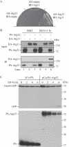
Figure 4. Interaction between Atg32 and Atg11
(A) Yeast two-hybrid analysis between Atg32 and Atg11. The wild-type (PJ69-4A) strain was transformed with AD-Atg32, BD-Atg11, or empty vectors as indicated. After selection on plates lacking uracil and leucine, the transformants were inoculated on an SMD plate lacking histidine and grown at 30°C for 4 d. (B) Atg32 associated with Atg11 during starvation. The atg32Δ strain transformed with a plasmid expressing protein A-tagged Atg32 or HA-tagged Atg11 under the control of the CUP1 promoter (pCuPA-Atg32 or pCuHA-Atg11) only, or both plasmids together was cultured in SMD medium to mid-log phase or starved in SD-N for 1 h. IgG sepharose was used to precipitate PA-Atg32 from cell lysates. The upper two panels show the immunoblot of total cell lysates (input, equivalent to A600 = 0.08 unit of cells were loaded) and the lower two panels show the IgG precipitates (IP, equivalent to A600 = 2.50 units of cells were loaded), which were probed with anti-HA and anti-protein A antibody. (C) A strain expressing Om45-GFP (TKYM22) transformed with a vector expressing protein A only (PA) or PA-Atg32 was cultured in SMGal medium to the mid-log growth phase, then shifted to SD-N medium for 0, 2, 4 and 6 h. The level of GFP processing, and the amount of PA and PA-Atg32 were monitored by immunoblotting with anti-YFP antibody and anti-PA antibody, respectively. The positions of molecular mass markers are indicated.
Figure 5
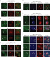
Figure 5. Mitochondria _target to the vacuole through a Cvt pathway-independent mechanism
The (A) atg11Δ, (B, D-F) atg1Δ, and (C) pep4Δ strains transformed with a plasmid expressing (A-C) GFP-tagged Atg32 under the control of the CUP1 promoter (pCuGFP-Atg32) and (D) pCFP-Ape1, (E) pCFP-Atg8, or (F) pHA-CFP-Atg11 were cultured in SMD medium, then shifted to starvation medium (SD-N) for 1.5 or 2 h. Cells were labeled with the vacuolar marker FM 4-64 and analyzed by fluorescence microscopy. The arrowheads in (B) and (D) mark GFP-Atg32, and the arrows mark non-overlapping CFP-Ape1 (D). The arrowheads and arrows in (E) and (F) mark overlapping and non-overlapping GFP-Atg32 and CFP-Atg8 or CFP-Atg11 puncta, respectively.
Figure 6
Figure 6. Atg32 localizes on mitochondria and _targets to the vacuole during mitophagy
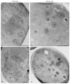
Cells expressing GFP-Atg32 under the GAL1 promoter were cultured in YPGal to mid-log phase (A and B), then shifted to SD-N for 4 h (C and D), prepared for electron microscopy using freeze substitution and stained with anti-YFP antibody followed by immunogold as described in Experimental Procedures. The arrow marks an example of a mitochondrion that is contained within a double-membrane autophagosome. AB, autophagic body; AP, autophagosome; M, mitochondria; V, vacuole; N, nucleus. Scale bar, 500 nm.
Figure 7
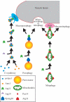
Figure 7. Model of selective autophagy
The cargo of the Cvt pathway, prApe1 and Ams1, form a complex with the receptor protein Atg19. Atg11 interacts with Atg19 to recruit the prApe1-Ams1-Atg19 complex to the PAS, the site of Cvt vesicle formation. When pexophagy is induced in Pichia pastoris, PpAtg30 binds the peroxisomal protein PpPex14. PpAtg30 further interacts with PpAtg11, allowing recruitment of the peroxisome to the PAS for both macropexophagy and micropexophagy. When mitophagy is induced, the mitochondrial protein Atg32 binds Atg11. Atg11 recruits mitochondria to the vacuolar surface, where uptake may occur via the PAS (through a macroautophagy-like process) or independent of this structure (through a microautophagy-like process).

Comment in

Similar articles

Cited by

References

    1. Abeliovich H, Zhang C, Dunn WA, Jr., Shokat KM, Klionsky DJ. Chemical genetic analysis of Apg1 reveals a non-kinase role in the induction of autophagy. Mol Biol Cell. 2003;14:477–490. - PMC - PubMed
    1. Baba M. Electron microscopy in yeast. Methods Enzymol. 2008;451:133–149. - PubMed
    1. Bogenhagen DF. Repair of mtDNA in vertebrates. Am J Hum Genet. 1999;64:1276–1281. - PMC - PubMed
    1. Cao Y, Klionsky DJ. Physiological functions of Atg6/Beclin 1: a unique autophagy-related protein. Cell Res. 2007;17:839–849. - PubMed
    1. Cheong H, Nair U, Geng J, Klionsky DJ. The Atg1 kinase complex is involved in the regulation of protein recruitment to initiate sequestering vesicle formation for nonspecific autophagy in Saccharomyces cerevisiae. Mol Biol Cell. 2008;19:668–681. - PMC - PubMed

Publication types

MeSH terms

  NODES
twitter 2