. 2009 Oct;183(2):581-94.
doi: 10.1534/genetics.109.103945. Epub 2009 Jul 27.

The role of MITF phosphorylation sites during coat color and eye development in mice analyzed by bacterial artificial chromosome transgene rescue

Affiliations

The role of MITF phosphorylation sites during coat color and eye development in mice analyzed by bacterial artificial chromosome transgene rescue

Georg L Bauer et al. Genetics. 2009 Oct.

Abstract

The microphthalmia-associated transcription factor (Mitf) has emerged as an important model for gene regulation in eukaryotic organisms. In vertebrates, it regulates the development of several cell types including melanocytes and has also been shown to play an important role in melanoma. In vitro, the activity of MITF is regulated by multiple signaling pathways, including the KITL/KIT/B-Raf pathway, which results in phosphorylation of MITF on serine residues 73 and 409. However, the precise role of signaling to MITF in vivo remains largely unknown. Here, we use a BAC transgene rescue approach to introduce specific mutations in MITF to study the importance of specific phospho-acceptor sites and protein domains. We show that mice that carry a BAC transgene where single-amino-acid substitutions have been made in the Mitf gene rescue the phenotype of the loss-of-function mutations in Mitf. This may indicate that signaling from KIT to MITF affects other phospho-acceptor sites in MITF or that alternative sites can be phosphorylated when Ser73 and Ser409 have been mutated. Our results have implications for understanding signaling to transcription factors. Furthermore, as MITF and signaling mechanisms have been shown to play an important role in melanomas, our findings may lead to novel insights into this resilient disease.


Figures

F<sc>igure</sc> 1.—
Figure 1.—
The BAC clones and recombineering strategy. (A) The two BAC clones isolated in the study are shown with respect to the structure of the Mitf gene. The size of the two clones are indicated in parentheses. Also indicated is the distance from the ends of the first and last known exons of Mitf to the ends of each clone. BAC Mi1 lacks 14.2 kb of DNA with respect to the first nucleotide of exon 1A whereas it contains an additional 47.5 kb of DNA from the last known nucleotide of exon 9. Similarly, BAC Mi2 lacks 76 kb with respect to the first nucleotide of exon 1A and contains additional 93.9 kb at the 3′-end. The relative sizes of the exons and introns are indicated but are not shown in the correct proportional sizes. The locations of amino acid residues Ser73 and Ser409 are indicated. (B) The two-step recombineering strategy used for generating the Ser73 and Ser409 mutations. The hit oligo was recombineered into the Mitf locus, and successful recombinants were screened using Hit-specific primers. After verifying the recombineered Hit clone, the Fix oligo was recombineered into the positive Hit-BAC and screened by PCR using fix-specific oligos. The resulting mutated BAC clones were further screened for the presence of the mutation and the absence of the Hit sequences before they were used for generating transgenic mice. (C) BAC transgene rescue of the Mitfmi-ew mutation. All three mice are homozygous for the Mitfmi-ew mutation, and therefore all should have the phenotype of the mouse on the left showing a white coat and microphthalmia. However, the two mice on the right also carry the BAC Mi1 clone, which fully rescues both phenotypes.
F<sc>igure</sc> 2.—
Figure 2.—
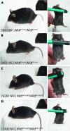
Wild-type BAC clones rescue Mitf mutant phenotypes. The two wild-type BAC clones rescue Mitf mutant phenotypes completely. (A and B) The BAC Mi1 and Mi2 wild-type BAC clones rescue the phenotype of the Mitfmi-ew mutation fully; no belly spot is visible in the mice (right panels). (C and D) The BAC Mi1 clone also rescues the Mitfmi-vga9 loss-of-function phenotype fully as seen in two independent transgenic lines. Again, not even a belly spot is visible, suggesting full rescue. The inserts show the eyes of the BAC transgenic mice.
F<sc>igure</sc> 3.—
Figure 3.—
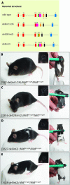
The mutant BAC clones Ser73Ala and Ser409Ala rescue the Mitf mutant phenotype. The BAC clones containing the Ser73Ala and Ser409Ala mutations rescue the phenotype of the Mitfmi-vga9 loss-of-function mutation fully in either single or double-mutant combination. (A–C) Three different transgene lines that contain the Ser73Ala mutation were obtained. The line 21055 fully rescues the microphthalmia associated with the Mitfmi-vga9 mutation whereas the coat color phenotype is rescued only partially (A). These animals lack pigmentation on the belly and have small unpigmented spots over the rest of the body. However, a significant proportion of the coat is pigmented, suggesting significant rescue in this line; the spots on the back are rather small, suggesting failure to rescue during late melanocyte development or differentiation. (B) The line 8260 rescues the phenotype fully, apart from a small belly spot (right). (C) The line 8250 rescues the coat color phenotype fully except for a minor belly streak seen in most of the animals. However, eye development is not rescued. The phenotypic differences in these three lines are most likely due to differences in the integration event of the transgenes. (D) The Ser409Ala mutant BAC clone can rescue the phenotype of the Mitfmi-vga9 mutation completely, although many animals show a significant belly spot. (E) The BAC clone containing the double mutation Ser73Ala; Ser409Ala can also rescue the phenotype of the Mitfmi-vga9 mutation completely, and in this case belly spots are rarely seen. The inserts show the eyes of the BAC transgenic mice.
F<sc>igure</sc> 4.—
Figure 4.—
Splicing of exon 2 of Mitf is affected in the BAC transgenic mice containing the Ser73Ala mutation. RT–PCR was used to determine if splicing of exon 2 was affected in the BAC transgenic mice. Primers spanning from exons 1B to exon 3 (bottom) were used to amplify cDNAs isolated from the indicated BAC transgenic mice (top). No products were amplified in the homozygous Mitfmi-vga9 mice whereas two bands were amplified in Mitfmi-vga9 heterozygotes and in wild-type mice as well as in all the other BAC transgenic lines. The 359-bp top band represents the full-length product whereas the 191-bp bottom band represents an alternative product lacking exon 2B. Quantitative PCR using exon-specific primers was used to determine the relative amount of these two products in the different BAC transgenic lines. The results are indicated as the percentage of the product lacking exon 2B over the total amount of products lacking and containing exon 2B. The shorter product is relatively more abundant in the lines that contain the Ser73Ala mutation.
F<sc>igure</sc> 5.—
Figure 5.—
Generation and analysis of exon 2 deletion mutations. Several different deletion mutations were generated to analyze the role of exons 2A and B in Mitf function. (A) The genomic structure of the deletion mutations that were generated. The exons are indicated as colored boxes. (B–E) The transgenic mice carrying the different deletion mutations. The left panels show a side view of the mice and the right panels show the belly region. The inserts in the upper right corner of the left panels show the eyes of the BAC transgenic mice. (B) The BAC carrying the del(int1/2A) mutation rescues the phenotype of the Mitfmi-vga9 mutation to a large extent, leaving a sizable belly spot. (C) The BAC carrying the del(2B/int2) mutation also rescues the phenotype of the Mitfmi-vga9 mutation; a small belly spot is visible in most of the animals. (D and E) Two different transgenic lines were generated which carry the BAC with the del(int2) mutation. These lines fully rescue the microphthalmia associated with the Mitfmi-vga9 mutation whereas coat color is rescued only partially. These animals have an extensive belly spot in addition to white spots over the rest of the coat.
F<sc>igure</sc> 6.—
Figure 6.—
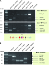
Analysis of splicing in BAC transgenic mice carrying Mitf deletion mutations. RT–PCR analysis was performed on both heart and skin to characterize Mitf transcripts in mice carrying the BAC deletion mutations. (A) Splicing of Mitf in heart tissue was characterized using two primer sets. The first primer set (top) extended from exon 1B to exon 3. In wild-type mice, these primers result in two bands: a full-length 359-bp band and a shorter 191-bp band lacking exon 2B. The full-length band is more prominent, representing 90–95% of the product. In the del(int1/2A) mutation, a single 131-bp PCR product was made, representing a transcript lacking both exons 2A and 2B. Because exon 2A is missing, the only product possible is made by splicing exon 1B directly to exon 3. In the del(2B/int2) mutation, only the 191-bp product lacking exon 2B is generated, as expected. The two del(int2) lines generate only the full-length product. The Mitfmi-enu22(398) mice express both the full-length product and the 131-bp product missing both exons 2A and 2B. The second primer pair (bottom) extended from exon 1B to exon 4. The results are largely the same as above. The only exception is that the two del(int2) lines produce a smaller band in addition to the full-length product observed in line TG13628. The smaller product represents transcripts that lack exons 2B and 3, suggesting that exon 2B is spliced directly to exon 4 in these mice. (B) Splicing of Mitf in skin was analyzed using primers extending from exon 1M to exon 4. In control (B6) animals, this primer set results in the production of a single 440-bp full-length product. These primers result in two products in skin from mice carrying the del(int1/2A) deletion mutation, a 380-bp band representing a transcript lacking exon 2A, and a smaller 212-bp band lacking exons 2A and 2B. This deletion fuses exon 1M directly to exon 2B so none of the products will contain exon 2A. However, the smaller product suggests that the splice donor in exon 1M is still active and that this exon can be fused directly to exon 3. The del(2B/int2) deletion also results in the production of two fragments using this primer set: a 272-bp fragment lacking exon 2B and a fragment lacking exons 2A, 2B, and 3. In these mice, exon 2A is fused directly to exon 3 so all products will lack exon 2B. The small alternative product indicates that exon 1M can also be spliced directly to exon 4. Both del(int2) lines result in only one product, representing a transcript lacking exons 2B and 3. Clearly, the absence of intron 2 leads to the skipping of the two flanking exons, and the only product produced fuses exon 2A directly to exon 4.
F<sc>igure</sc> 7.—
Figure 7.—
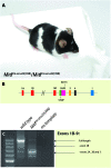
Molecular and phenotypic characterization of the Mitfmi-enu22(398) mutation. (A) The phenotype of the Mitfmi-enu22(398) mutation. The mice have normal eye size. However, although they have extensive white belly spots and white spots over the rest of the body, a significant portion of their coat is normally pigmented. (B) The mutation associated with this phenotype is a stop codon in exon 2A of the Mitf gene. The position of the Ser73 amino acid is also indicated. (C) Characterization of splicing of the Mitf gene in normal and Mitfmi-enu22(398) mutant mice using primers extending from exon 1B to exon 9. In normal mice, this results in the production of a major 997-bp full-length band and several alternative splice products, including the 829-bp product lacking exon 2B. In the Mitfmi-enu22(398) mutation, the major 685-bp product is lacking exons 2A, 2B, and 3.

Similar articles

Cited by

References

    1. Arnheiter, H., 2007. Mammalian paramutation: A tail's tale? Pigment Cell Res. 20: 36–40. - PMC - PubMed
    1. Bharti, K., J. Debbache, J. Wang and H. Arnheiter, 2009. The basic helix-loop-helix leucine-zipper gene Mitf: analysis of alternative promoter choice and splicing in Methods in Molecular Biology: Transcription Factors, edited by Paul Higgins. Humana Press, Totowa, NJ (in press). - PubMed
    1. Bismuth, K., S. Skuntz, J. H. Hallsson, E. Pak, A. S. Dutra et al., 2008. An unstable _targeted allele of the mouse Mitf gene with a high somatic and germline reversion rate. Genetics 178: 259–272. - PMC - PubMed
    1. Dubreuil, P., L. Forrester, R. Rottapel, M. Reedijk, J. Fujita et al., 1991. The c-fms gene complements the mitogenic defect in mast cells derived from mutant W mice but not mi (microphthalmia) mice. Proc. Natl. Acad. Sci. USA 88: 2341–2345. - PMC - PubMed
    1. Frame, S., and P. Cohen, 2001. GSK3 takes centre stage more than 20 years after its discovery. Biochem. J. 359: 1–16. - PMC - PubMed

Publication types

MeSH terms

Substances

  NODES
twitter 2
Verify 1