. 2009 Oct 6;106(40):17049-54.
doi: 10.1073/pnas.0903316106. Epub 2009 Sep 21.

The Tor and PKA signaling pathways independently _target the Atg1/Atg13 protein kinase complex to control autophagy

Affiliations

The Tor and PKA signaling pathways independently _target the Atg1/Atg13 protein kinase complex to control autophagy

Joseph S Stephan et al. Proc Natl Acad Sci U S A. .

Abstract

Macroautophagy (or autophagy) is a conserved degradative pathway that has been implicated in a number of biological processes, including organismal aging, innate immunity, and the progression of human cancers. This pathway was initially identified as a cellular response to nutrient deprivation and is essential for cell survival during these periods of starvation. Autophagy is highly regulated and is under the control of a number of signaling pathways, including the Tor pathway, that coordinate cell growth with nutrient availability. These pathways appear to _target a complex of proteins that contains the Atg1 protein kinase. The data here show that autophagy in Saccharomyces cerevisiae is also controlled by the cAMP-dependent protein kinase (PKA) pathway. Elevated levels of PKA activity inhibited autophagy and inactivation of the PKA pathway was sufficient to induce a robust autophagy response. We show that in addition to Atg1, PKA directly phosphorylates Atg13, a conserved regulator of Atg1 kinase activity. This phosphorylation regulates Atg13 localization to the preautophagosomal structure, the nucleation site from which autophagy pathway transport intermediates are formed. Atg13 is also phosphorylated in a Tor-dependent manner, but these modifications appear to occur at positions distinct from the PKA phosphorylation sites identified here. In all, our data indicate that the PKA and Tor pathways function independently to control autophagy in S. cerevisiae, and that the Atg1/Atg13 kinase complex is a key site of signal integration within this degradative pathway.


Conflict of interest statement

The authors declare no conflict of interest.

Figures
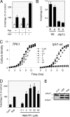
Fig. 1.
Fig. 1.
Inactivation of the Ras/PKA signaling pathway was sufficient to induce autophagy. (A) Autophagy induction upon rapamycin treatment and/or expression of the dominant-negative RAS2ala22 allele. Wild-type cells (TN125) were grown to mid-log phase and treated with 20 ng/mL rapamycin for 4 h at 30 °C. Cells carrying the MET3-RAS2ala22 allele were transferred to an SC minimal medium lacking methionine for 4 h at 30 °C to induce expression from the MET3 promoter. Autophagy levels were assessed with an alkaline phosphatase (ALP)-based assay as described in the Materials and Methods. The white bars indicate the relative levels of ALP activity in the untreated controls and the black bars the activity following the indicated treatments. (B) The autophagy activity induced upon inactivation of the Ras/PKA pathway was dependent upon the presence of the Atg1 protein. Autophagy levels were assessed with the ALP-based assay in isogenic wild-type and atg1Δ cells after 4 h of rapamycin treatment (R) or exposure to the RAS2ala22 protein (A). (C) Growth curves for isogenic TPK1 and tpk1-as strains in YPAD at 30 °C are shown. The drug 1NM-PP1 was present at the indicated concentrations. (D) Inactivation of PKA signaling in the tpk1-as strain resulted in the induction of autophagy. Autophagy levels were assessed with the ALP-based assay in tpk1-as cells (PHY4710) that were treated for 5 h with either 200 ng/mL rapamycin or the indicated concentrations of 1NM-PP1. (E) Cells over-expressing Ape1 were treated for 4 h with either 200 ng/ml rapamycin or 25 μM 1NM-PP1, and the relative level of Ape1 processing was assessed by Western blotting. Cont, untreated cells.
Fig. 2.
Fig. 2.
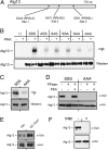
The Atg13 protein was a substrate for PKA. (A) The three conserved sites of PKA phosphorylation in Atg13 are shown. (B and C) The in vitro phosphorylation of Atg13 was dependent upon the presence of the above three PKA sites. The indicated Atg13 variants were precipitated from yeast cells and incubated with [γ-32P] ATP and either bovine PKA (bPKA) (in B) or the S. cerevisiae Tpk1 (C). S, serine; A, alanine. (D) The three PKA sites were required for the in vivo phosphorylation of Atg13. The indicated Atg13 proteins were immunoprecipitated from yeast cell extracts with an α-HA antibody and treated with λ phosphatase and then incubated with bPKA and 3 mM ATP, as indicated. The level of PKA phosphorylation was assessed by Western blotting with an α-substrate antibody that recognizes phosphorylated PKA sites. (E) The in vivo level of PKA phosphorylation on Atg13 was elevated in a strain over-expressing Tpk1. (F) Recognition by the α-substrate antibody was lost following inactivation of PKA. The tpk1-as strain was incubated with 10 μM 1NM-PP1 for 4 h and the PKA phosphorylation level of Atg13 was assessed by Western blotting with the α-substrate antibody.
Fig. 3.
Fig. 3.
The loss of PKA phosphorylation on Atg13 preceded the activation of Atg1 and the induction of autophagy. (A) Atg13 and Atg17 were required for Atg1 autophosphorylation in vivo. Cell extracts were prepared from the indicated yeast strains and the levels of autophosphorylated Atg1 were assessed by Western blotting. The cells were treated with 200 ng/mL rapamycin for 0 or 2 h at 30 °C. The atg1KD strain has a kinase-defective allele of ATG1, atg1-K54A. In the bottom panel, Atg1 was immunoprecipitated from yeast cell extracts and then treated with λ phosphatase, as indicated. (B) The relative levels of Atg13 phosphorylation by PKA (top panel) and of the “activated” form of Atg1 (bottom) were assessed by Western blotting at the indicated times after the addition of 25 μM 1NM-PP1 to a culture of tpk1-as cells. (C) Autophagy activity was assessed with the ALP-based assay at the indicated times after the addition of 25 μM 1NM-PP1 to tpk1-as cells. (D) The localization of an Atg1-YFP protein was assessed by fluorescence microscopy 4 h after the addition of 25 μM 1NM-PP1 to a culture of tpk1-as cells.
Fig. 4.
Fig. 4.
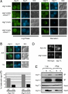
The PAS localization of Atg13 was regulated by PKA phosphorylation. (A) Fluorescence microscopy was performed with cells that contained the indicated YFP-Atg13 fusion proteins. The Atg13 proteins had either wild-type (Atg13-SSS) or nonphosphorylatable versions (Atg13-AAA) of the three PKA sites. The CFP-Atg11 fusion protein was present in all cells and served as a marker for the PAS. The RAS2val19 allele was present in the indicated strains. Nitrogen starvation was achieved by transferring the cells from SC glucose minimal medium to the SD-N medium for 1 h at 30 °C. (B) The localization of a wild-type Atg13-YFP protein in the indicated cells was assessed by fluorescence microscopy. A22, RAS2ala22. (C) Atg17 was required for the efficient localization of Atg13-AAA to the PAS in log phase cells. The fraction of cells with Atg1-AA or Atg13-AAA present in a perivacuolar punctate spot in the indicated strains is shown (20). At least 50 cell images were examined for each strain. (D) Representative images for the indicated strains in C. (E) Alteration of the PKA phosphorylation sites in Atg13 influenced the interaction with Atg17. The indicated Atg13 proteins were immunoprecipitated from yeast cell extracts and the relative levels of the associated Atg17 were assessed by Western blotting. The cell extracts were prepared from either mid-log phase cultures (Log) or from cells that were treated with rapamycin (Rap). S, Atg13-SSS; A, Atg13-AAA.
Fig. 5.
Fig. 5.
The PKA and Tor pathways independently _target Atg13 to control autophagy activity. (A) Simultaneous inactivation of the PKA and Tor pathways resulted in an elevated autophagy response relative to the loss of either pathway alone. Autophagy levels were assessed with the ALP-based assay in tpk1-as cells that had been treated for the indicated time with either 25 μM 1NM-PP1, 200 ng/mL rapamycin, or both reagents. The data shown are from a single experiment that was representative of at least three independent replicates. (B) The PKA phosphorylation of Atg13 was not diminished upon inactivation of the Tor pathway. Atg13 was immunoprecipitated from cells following a 2 h treatment with either 20 (Lo) or 200 (Hi) ng/mL rapamycin. The level of PKA phosphorylation was subsequently assessed by Western blotting with the α-PKA substrate antibody. (C) Inactivation of the Ras/PKA pathway did not influence the Tor-dependent phosphorylation of Atg13. Cell extracts were prepared from the indicated cells and the level of Tor-dependent phosphorylation of Atg13 was assessed by Western blotting. In the left-hand lanes, cells containing either MET3-RAS2ala22 (A22) or a control plasmid (-) were incubated in methionine-free medium for 6 h to allow for expression from the MET3 promoter. In the middle lanes, the tpk1-as strain, PHY4710, was incubated for 4 h with 0 or 5 μM 1NM-PP1. The bottom panel in the middle lanes shows the level of PKA phosphorylation on Atg13 as assessed by Western blotting with the α-PKA substrate antibody. Note that the relative spread of the Atg13 “smear” is dependent upon the running conditions of the gel. (D) A model depicting the proposed roles of the PKA and Tor pathways in the control of autophagy. The dashed lines indicate the Atg1 and Atg13 interactions with each other or the PAS (and perhaps Atg17), and the solid lines indicate the regulatory effects of the PKA and/or Tor pathways on these interactions. See the text for additional details.

Similar articles

Cited by

References

    1. Noda T, Suzuki K, Ohsumi Y. Yeast autophagosomes: De novo formation of a membrane structure. Trends Cell Biol. 2002;12:231–235. - PubMed
    1. Xie Z, Klionsky DJ. Autophagosome formation: Core machinery and adaptations. Nat Cell Biol. 2007;9:1102–1109. - PubMed
    1. Kopitz J, Kisen GO, Gordon PB, Bohley P, Seglen PO. Nonselective autophagy of cytosolic enzymes by isolated rat hepatocytes. J Cell Biol. 1990;111:941–953. - PMC - PubMed
    1. Schworer CM, Mortimore GE. Glucagon-induced autophagy and proteolysis in rat liver: Mediation by selective deprivation of intracellular amino acids. Proc Natl Acad Sci USA. 1979;76:3169–3173. - PMC - PubMed
    1. Levine B, Klionsky DJ. Development by self-digestion: Molecular mechanisms and biological functions of autophagy. Dev Cell. 2004;6:463–477. - PubMed

Publication types

MeSH terms

Substances

  NODES
Note 1
twitter 2