Increased muscle PGC-1alpha expression protects from sarcopenia and metabolic disease during aging
- PMID: 19918075
- PMCID: PMC2787152
- DOI: 10.1073/pnas.0911570106
Increased muscle PGC-1alpha expression protects from sarcopenia and metabolic disease during aging
Erratum in
- Proc Natl Acad Sci U S A. 2014 Nov 4;111(44):15851
Retraction in
-
Retraction for Wenz et al., Increased muscle PGC-1α expression protects from sarcopenia and metabolic disease during aging.Proc Natl Acad Sci U S A. 2016 Dec 27;113(52):E8502. doi: 10.1073/pnas.1619713114. Epub 2016 Dec 19. Proc Natl Acad Sci U S A. 2016. PMID: 27994157 Free PMC article. No abstract available.
Abstract
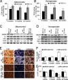
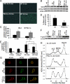
Aging is a major risk factor for metabolic disease and loss of skeletal muscle mass and strength, a condition known as sarcopenia. Both conditions present a major health burden to the elderly population. Here, we analyzed the effect of mildly increased PGC-1alpha expression in skeletal muscle during aging. We found that transgenic MCK-PGC-1alpha animals had preserved mitochondrial function, neuromuscular junctions, and muscle integrity during aging. Increased PGC-1alpha levels in skeletal muscle prevented muscle wasting by reducing apoptosis, autophagy, and proteasome degradation. The preservation of muscle integrity and function in MCK-PGC-1alpha animals resulted in significantly improved whole-body health; both the loss of bone mineral density and the increase of systemic chronic inflammation, observed during normal aging, were prevented. Importantly, MCK-PGC-1alpha animals also showed improved metabolic responses as evident by increased insulin sensitivity and insulin signaling in aged mice. Our results highlight the importance of intact muscle function and metabolism for whole-body homeostasis and indicate that modulation of PGC-1alpha levels in skeletal muscle presents an avenue for the prevention and treatment of a group of age-related disorders.
Conflict of interest statement
The authors declare no conflict of interest.
Figures


Similar articles
-
alpha-Lipoic acid increases energy expenditure by enhancing adenosine monophosphate-activated protein kinase-peroxisome proliferator-activated receptor-gamma coactivator-1alpha signaling in the skeletal muscle of aged mice.Metabolism. 2010 Jul;59(7):967-76. doi: 10.1016/j.metabol.2009.10.018. Epub 2009 Dec 16. Metabolism. 2010. PMID: 20015518 Free PMC article.
-
Functional effects of muscle PGC-1alpha in aged animals.Skelet Muscle. 2020 May 6;10(1):14. doi: 10.1186/s13395-020-00231-8. Skelet Muscle. 2020. PMID: 32375875 Free PMC article.
-
Skeletal muscle-specific expression of PGC-1α-b, an exercise-responsive isoform, increases exercise capacity and peak oxygen uptake.PLoS One. 2011;6(12):e28290. doi: 10.1371/journal.pone.0028290. Epub 2011 Dec 8. PLoS One. 2011. PMID: 22174785 Free PMC article.
-
Role of PGC-1α in sarcopenia: etiology and potential intervention - a mini-review.Gerontology. 2015;61(2):139-48. doi: 10.1159/000365947. Epub 2014 Dec 6. Gerontology. 2015. PMID: 25502801 Review.
-
PGC-1alpha-induced improvements in skeletal muscle metabolism and insulin sensitivity.Appl Physiol Nutr Metab. 2009 Jun;34(3):307-14. doi: 10.1139/H09-008. Appl Physiol Nutr Metab. 2009. PMID: 19448691 Review.
Cited by
-
Mitochondrial Diseases Part III: Therapeutic interventions in mouse models of OXPHOS deficiencies.Mitochondrion. 2015 Jul;23:71-80. doi: 10.1016/j.mito.2015.01.007. Epub 2015 Jan 29. Mitochondrion. 2015. PMID: 25638392 Free PMC article. Review.
-
Sarcopenia and cachexia: the adaptations of negative regulators of skeletal muscle mass.J Cachexia Sarcopenia Muscle. 2012 Jun;3(2):77-94. doi: 10.1007/s13539-011-0052-4. Epub 2012 Jan 12. J Cachexia Sarcopenia Muscle. 2012. PMID: 22476916 Free PMC article.
-
Attenuated Oxidative Stress following Acute Exhaustive Swimming Exercise Was Accompanied with Modified Gene Expression Profiles of Apoptosis in the Skeletal Muscle of Mice.Oxid Med Cell Longev. 2016;2016:8381242. doi: 10.1155/2016/8381242. Epub 2016 Apr 10. Oxid Med Cell Longev. 2016. PMID: 27143996 Free PMC article.
-
Oleic acid stimulates complete oxidation of fatty acids through protein kinase A-dependent activation of SIRT1-PGC1α complex.J Biol Chem. 2013 Mar 8;288(10):7117-26. doi: 10.1074/jbc.M112.415729. Epub 2013 Jan 17. J Biol Chem. 2013. PMID: 23329830 Free PMC article.
-
Akt1-mediated skeletal muscle growth attenuates cardiac dysfunction and remodeling after experimental myocardial infarction.Circ Heart Fail. 2012 Jan;5(1):116-25. doi: 10.1161/CIRCHEARTFAILURE.111.964783. Epub 2011 Dec 1. Circ Heart Fail. 2012. PMID: 22135402 Free PMC article.
References
-
- Dela F, Kjaer M. Resistance training, insulin sensitivity and muscle function in the elderly. Essays Biochem. 2006;42:75–88. - PubMed
-
- Marcell TJ. Sarcopenia: Causes, consequences, and preventions. J Gerontol A Biol Sci Med Sci. 2003;58:M911–M916. - PubMed
-
- Taaffe DR. Sarcopenia—Exercise as a treatment strategy. Aust Fam Physician. 2006;35:130–134. - PubMed
Publication types
MeSH terms
Substances
Grants and funding
- R01 DK054477/DK/NIDDK NIH HHS/United States
- R01 DK054477-11/DK/NIDDK NIH HHS/United States
- CA85700/CA/NCI NIH HHS/United States
- R56 NS041777/NS/NINDS NIH HHS/United States
- R01 NS041777/NS/NINDS NIH HHS/United States
- EY10804/EY/NEI NIH HHS/United States
- R01 EY010804/EY/NEI NIH HHS/United States
- AG0005917/AG/NIA NIH HHS/United States
- R01 DK061562/DK/NIDDK NIH HHS/United States
- R56 DK054477/DK/NIDDK NIH HHS/United States
- DK61562/DK/NIDDK NIH HHS/United States
- R01 CA085700/CA/NCI NIH HHS/United States
- NS041777/NS/NINDS NIH HHS/United States
- DK54477/DK/NIDDK NIH HHS/United States
- R01 AG005917/AG/NIA NIH HHS/United States
LinkOut - more resources
Full Text Sources
Other Literature Sources
Medical
Molecular Biology Databases

