. 2009 Nov 25;139(5):891-906.
doi: 10.1016/j.cell.2009.10.027.

Matrix crosslinking forces tumor progression by enhancing integrin signaling

Affiliations

Matrix crosslinking forces tumor progression by enhancing integrin signaling

Kandice R Levental et al. Cell. .

Abstract

Tumors are characterized by extracellular matrix (ECM) remodeling and stiffening. The importance of ECM remodeling to cancer is appreciated; the relevance of stiffening is less clear. We found that breast tumorigenesis is accompanied by collagen crosslinking, ECM stiffening, and increased focal adhesions. Induction of collagen crosslinking stiffened the ECM, promoted focal adhesions, enhanced PI3 kinase (PI3K) activity, and induced the invasion of an oncogene-initiated epithelium. Inhibition of integrin signaling repressed the invasion of a premalignant epithelium into a stiffened, crosslinked ECM and forced integrin clustering promoted focal adhesions, enhanced PI3K signaling, and induced the invasion of a premalignant epithelium. Consistently, reduction of lysyl oxidase-mediated collagen crosslinking prevented MMTV-Neu-induced fibrosis, decreased focal adhesions and PI3K activity, impeded malignancy, and lowered tumor incidence. These data show how collagen crosslinking can modulate tissue fibrosis and stiffness to force focal adhesions, growth factor signaling and breast malignancy.


Figures
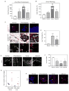
Figure 1
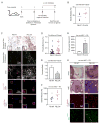
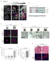
Figure 1. ECM stiffening, collagen crosslinking and tissue fibrosis accompany breast transformation
A. Elastic modulus of mammary glands from FVB MMTV-Neu mice at different stages of tumor progression measured by unconfined compression. B. Shear rheology of tissues described in A. C. Top row: Confocal images of tissues in A stained for cytokeratin 14 (red) and DAPI (nuclei; blue). Middle and bottom rows: Photomicrographs of tissues in A stained with Picrosirius Red and hematoxylin, viewed under parallel (middle), and orthogonal polarizing filters (bottom). D. Quantification of images in C. E. SHG images of tissues described in A. F. Quantification of images in E. G. Scatter plot of collagen crosslinks. H. Confocal images of tissues described in A. stained for lysyl oxidase (LOX; red) and DAPI (nuclei; blue). Images 40×, Bar 50μm. Values in A, B & D; Mean ± SEM of 4-6 glands/condition. * p ≤0.05, ** p≤0.01, *** p≤0.001.
Figure 2
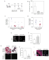
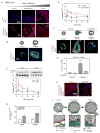
Figure 2. Collagen remodeling and tissue stiffening promote focal adhesions and tumor invasion
A. Experimental design. B. Scatter plot of breast rheology following LOX (FB.LOX) or control (FB.ctrl) fibroblast conditioning. C. Top two panels: Photomicrographs of tissues described in B stained with Picrosirius Red and hematoxylin viewed under parallel and orthogonal polarizing filters. Bar 50μm. Third panel: SHG images of tissues from fibroblast-conditioned mammary glands. Bar 25μm. Fourth and fifth panels: Confocal images of fibroblasts residing within fibroblast-conditioned mammary glands stained for active FAK (FAKpY397; red; fourth panel), p130Cas (red; fifth panel) and DAPI (nuclei; blue). Bar 50μm. D. Scatter plot of fibrillar collagen in tissue shown in C. E. Scatter plot of collagen linearity measured by Curvature Ratio from SHG images shown in C. F. Rheology of mammary glands with injected Ha-ras MCF10AT MECs. G. Lesion burden for mouse cohorts. H. Top: Photographs of mammary glands three weeks after injection with Ha-ras MCF10AT organoids. Middle: H&E stained tissue of MECs from lesions shown in H. Bottom: Confocal images of MECs in tissue shown in H stained for β1 integrin (red), active FAK (FAKpY397; green) and DAPI (nuclei; blue; insert). Bar 50μm. Values in E and G are Mean ± SEM of several glands. * p≤0.05.
Figure 3
Figure 3. Inhibiting collagen crosslinking tempers tissue fibrosis and reduces focal adhesions
A. Experimental design. B. LOX enzymatic activity in serum of untreated (Control) compared to BAPN (+BAPN) or LOX inhibitory antibody-treated (+LOX-Ab) Neu mice. Values: Mean ± SEM. C. Scatter plots of reducible collagen crosslinks in LOX-inhibited (LOX-Inhib) and untreated (Control) Neu breasts. D. Scatter plots of pyridinoline in LOX-inhibited (LOX-Inhib) and untreated (Control) Neu breasts. E. SHG images of mammary glands from BAPN treated animals as described above. Bar 25μm. F. Collagen curvature in SHG images from D. (Supp Methods). Mean ± SEM 3-5 regions/4 glands/condition. G. Photomicrographs of sections from control and BAPN treated (LOX-Inhib) Neu mammary glands stained with Picrosirius Red and hematoxylin viewed under parallel or orthogonal polarizing filters. Bar 50μm. H. Fibrillar collagen in control and LOX inhibited glands from images shown in F. Mean ± SEM of 4-6 images/4-8 glands/condition. I. Confocal images of sections from control and BAPN treated Neu glands stained for active FAK (FAKpY397; red) and DAPI (nuclei; blue). Bar 50μm.* p ≤0.05, ** p≤0.01, *** p≤0.001.
Figure 4
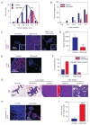
Figure 4. Inhibiting crosslinking reduces tissue fibrosis and tumor incidence and enhances tumor latency
A. Tumor latency. B. Tumor incidence without (Control) or with LOX inhibition (LOX-Inhib). C. Confocal images of activate ErbB2 (ErbB2pY1248; red) in tissues from FVB non transgenic littermates, Neu (MMTV-Neu) and LOX inhibited mice (MMTV-Neu +LOX-Inhib). Bar 50μm. D. Tumor size in control and LOX inhibited mice. E. Confocal images of sections from LOX inhibited and control glands stained for PCNA (red) and DAPI (nuclei; blue). Bar 50μm. F. Grading of tumor lesions from mammary glands of control (n=14) and BAPN treated (LOX-Inhib, n=13) animals. H. H&E stained tissue showing typical Normal Glandular structure, Hyperplastic alveolar nodules (HAN) and mammary intraepithelial neoplasia (MIN) premalignant lesions and Grades I, II and III ductal tumors. I. Confocal images of breast tissue stained for cytokeratin 14 (red) and DAPI (nuclei; blue) in control and LOX inhibited tissue. Bar 50μm. I. Percent cytokeratin 14 positive glands detected in breast from control and LOX inhibited animals. Values in A, B, D, F & I; Mean ± SEM of 4-6 measurements/4-12 tissue sections/group. * p ≤ 0.05, ** p ≤ 0.01, *** p ≤ 0.001.
Figure 5
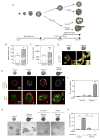
Figure 5. Collagen crosslinking and ECM stiffening promote focal adhesions and invasion of ErbB2 mammary colonies
A. Experimental design. B. Elastic modulus of ribose crosslinked (+Ribose) and untreated (Control) collagen gels. C. Cross sectional area of MEC colonies in +Ribose and Control gels. D. Confocal images of MCF10A colonies stained for β1 integrin (green), activate FAK (FAKpY397; red) and DAPI (nuclei; blue) in Control and +Ribose gels. Bar 20μm. E. Top panels: Confocal images of MEC colonies expressing the ErbB2 chimera stained for β catenin (green), β4 integrin (red) and DAPI (nuclei; blue) in Control or +Ribose gels with (+ErbB2) or without ErbB2 activity. Bottom panels: SHG images of collagen fibrils and confocal images of eGFP expressing MEC colonies as described above. Bar 20μm. Yellow arrows identify collagen bundles surrounding the colony periphery and white arrows indicate an invading MEC. F. Percent colony invasion shown in E. G. Phase contrast images of MEC colonies expressing the ErbB2 chimera in Control or +Ribose gels, with active ErbB2 (+ErbB2); co expressing a doxycycline inducible FRNK (right panel) or treated with β1 integrin function blocking antibody (+β1 block). H. Percent colony invasion shown in G. Values in B, C, F & H; Mean ± SEM from 3-5 experiments and/or 50-100 colonies in 3 experiments. ** p ≤ 0.01, *** p ≤ 0.001.
Figure 6
Figure 6. β1 integrin clustering promotes focal adhesions and drives invasion of a premalignant mammary epithelium in culture and in vivo
A. Combined and split (inserts) confocal images of tissue sections from MMTV-Neu breast stained for DAPI (nuclei; blue) and p130Cas (red; top), or β1 integrin (green; bottom) and, activate FAK (FAKpY397; red). Bar 20μm. (colocalization analysis of the malignant tumors: FAKpY397 and β1 integrin, Pearson's r = 0.78; p130Cas and β1 integrin, Pearson's r = 0.89) B. β1 integrin constructs used for the studies shown in C-G. C. Confocal images of MCF10A MEC rBM colonies expressing the β1 integrin wild type (β1(WT)) or clustering mutant (β1(V737N)) stained for β4 integrin (red; top), active FAK (FAKpY397; red; bottom) and DAPI (nuclei; blue). D. Phase contrast images of Ha-ras MCF10AT MEC colonies expressing the β1 integrin wild type, glycan wedge or integrin cluster mutant in rBM. Bar 50μm. E. Percent invasion of the colonies shown in D F. Lesion size formed by Ha-ras MCF10AT MECs expressing the clustering β1 integrin mutation (β1(V737N)) and wild type integrin (β1(WT)). G. Top panels: Photomicrographs of H&E stained sections of tumors formed by Ha-ras MECs expressing the clustering β1 integrin mutation (β1(V737N)) and wild type integrin (β1(WT)). Bottom panels: Confocal images of tissues stained for active FAK (FAKpY397; red) and DAPI (nuclei; blue). Bar 50μm. Values in E & F; Mean ± SEM; 12-50 measurements/3 experiments *** p≤ 0.001.
Figure 7
Figure 7. Tissue stiffness promotes integrin clustering and enhances growth factor-dependent PI3K activation
A. Confocal images of tissue from MMTV-Neu breast stained for Activated AktpS473 (red; top panels), active Akt Substrate (red; bottom panels) and DAPI (nuclei; blue). Bar 50μm. B. Confocal images of MEC colonies in untreated (Control) or ribose-crosslinked (+Ribose) collagen/rBM gels stained for active Akt substrate (green) and DAPI (nuclei; blue). Bar 50μm. C. Immunoblots and line graph of time course of AktpS473 and total Akt in EGF treated MCF10A MECs on soft and stiff rBM-PA gels. D. Ratio of AktpS473 to total Akt in MCF10A MECS with active (+ErbB2; Dox induced) and nonactive wild type ErbB2 (-ErbB2: non-induced) on soft and stiff rBM-PA gels. E. Time course of EGF activated AktpS473 to total Akt in MCF10A MECs expressing the wild type β1 integrin (β1(WT)) or the β1 integrin cluster mutant (β1(V737N)) on soft rBM-PA gels. F. Confocal images of MCF10A MEC colonies stained for β catenin (green), β4 integrin (red) and DAPI (nuclei; blue) in control or ribose-crosslinked collagen/rBM gels in the presence (+ErbB2) or absence of doxycycline; with or without the PI3K inhibitor, LY294002. Bar 20μm. G. Quantification of invasive colonies from F. H. Confocal images of sections from control and LOX inhibited Neu mice stained for active Akt substrate (red) and DAPI (nuclei, blue). Bar 50μm. I. Cartoon showing effect of ECM stiffness on mammary colony behavior. Values in C, D and E; Mean ± SEM of 3 experiments, 50-100 colonies/experiment. * p ≤ 0.05, ** p ≤ 0.01, *** p ≤ 0.001.

Comment in

  • Environment dictates behaviour.
    Baumann K. Baumann K. Nat Rev Mol Cell Biol. 2010 Oct;11(10):679. doi: 10.1038/nrm2984. Nat Rev Mol Cell Biol. 2010. PMID: 20861875 No abstract available.

Similar articles

Cited by

References

    1. Atsawasuwan P, Mochida Y, Katafuchi M, Kaku M, Fong KS, Csiszar K, Yamauchi M. Lysyl oxidase binds transforming growth factor-beta and regulates its signaling via amine oxidase activity. J Biol Chem. 2008;283:34229–34240. - PMC - PubMed
    1. Bierie B, Chung CH, Parker JS, Stover DG, Cheng N, Chytil A, Aakre M, Shyr Y, Moses HL. Abrogation of TGF-beta signaling enhances chemokine production and correlates with prognosis in human breast cancer. J Clin Invest. 2009;119:1571–1582. - PMC - PubMed
    1. Bierie B, Moses HL. Tumour microenvironment: TGFbeta: the molecular Jekyll and Hyde of cancer. Nat Rev Cancer. 2006;6:506–520. - PubMed
    1. Bondareva A, Downey CM, Ayres F, Liu W, Boyd SK, Hallgrimsson B, Jirik FR. The lysyl oxidase inhibitor, beta-aminopropionitrile, diminishes the metastatic colonization potential of circulating breast cancer cells. PLoS One. 2009;4:e5620. - PMC - PubMed
    1. Butcher DT, Alliston T, Weaver VM. A tense situation: forcing tumour progression. Nat Rev Cancer. 2009;9:108–122. - PMC - PubMed

Publication types

  NODES
twitter 2