. 2010 Jan 15;21(2):311-22.
doi: 10.1091/mbc.e09-05-0437. Epub 2009 Nov 25.

Nuclear-cytoplasmic shuttling of Chibby controls beta-catenin signaling

Affiliations

Nuclear-cytoplasmic shuttling of Chibby controls beta-catenin signaling

Feng-Qian Li et al. Mol Biol Cell. .

Abstract

In the canonical Wnt pathway, beta-catenin acts as a key coactivator that stimulates _target gene expression through interaction with Tcf/Lef transcription factors. Its nuclear accumulation is the hallmark of active Wnt signaling and is frequently associated with cancers. Chibby (Cby) is an evolutionarily conserved molecule that represses beta-catenin-dependent gene activation. Although Cby, in conjunction with 14-3-3 chaperones, controls beta-catenin distribution, its molecular nature remains largely unclear. Here, we provide compelling evidence that Cby harbors bona fide nuclear localization signal (NLS) and nuclear export signal (NES) motifs, and constitutively shuttles between the nucleus and cytoplasm. Efficient nuclear export of Cby requires a cooperative action of the intrinsic NES, 14-3-3, and the CRM1 nuclear export receptor. Notably, 14-3-3 docking provokes Cby binding to CRM1 while inhibiting its interaction with the nuclear import receptor importin-alpha, thereby promoting cytoplasmic compartmentalization of Cby at steady state. Importantly, the NLS- and NES-dependent shuttling of Cby modulates the dynamic intracellular localization of beta-catenin. In support of our model, short hairpin RNA-mediated knockdown of endogenous Cby results in nuclear accumulation of beta-catenin. Taken together, these findings unravel the molecular basis through which a combinatorial action of Cby and 14-3-3 proteins controls the dynamic nuclear-cytoplasmic trafficking of beta-catenin.


Figures

Figure 1.
Figure 1.
Cby harbors a functional NLS at the C-terminal end. (A) Cby contains two putative NLSs (NLS1 and NLS2). Cby NLS mutants generated by alanine substitutions are shown. CbyΔNLS2 lacks the last four C-terminal amino acids of the protein. (B) The C-terminal NLS2 is critical for nuclear import of Cby. Cos7 cells were transiently transfected with an expression vector encoding Flag-tagged at the N- or C-terminus (C-Flag) of WT or mutant Cby as indicated. Cells were fixed 24 h after transfection, followed by immunostaining with anti-Flag antibody. Nuclei were stained with DAPI. (C) Quantitative analysis of the results in B. The subcellular localization of Flag-tagged Cby was scored as follows: N>C, predominantly nuclear; N = C, evenly distributed between the nucleus and cytoplasm; N
Figure 2.
Figure 2.
Importin-α binds to Cby and facilitates its nuclear entry. (A) Cby interacts with multiple importin-α isoforms through the NLS2 motif. Bacterially expressed and purified MBP-CbyWT, MBP-CbyΔNLS2, or MBP-CbyR76A/R78A/R79A was incubated with GST fusion proteins with different importin-α members (1, 3, and 5–7). The complexes were then pulled down with amylose beads and subjected to Western blotting using anti-GST antibody. Input corresponds to the same amounts of fusion proteins used in each reaction mixture, visualized by silver staining to confirm that equal amounts of recombinant proteins were used. (B) Importin-β does not interact with Cby. In vitro pulldown assays were performed using GST-importin-β as described in A. Ten percent of each reaction mixture was resolved on a separate SDS-PAGE, followed by immunoblotting with anti-GST and anti-MBP antibodies to verify that equal amounts of recombinant proteins were used. (C) Overexpression of importin-α promotes nuclear localization of CbyWT but not that of CbyΔNLS2. Cos7 cells were cotransfected with an expression construct for Flag-tagged CbyWT or CbyΔNLS2 and a negative control empty vector or an HA-importin-α3 expression plasmid and immunostained with anti-Flag antibody.
Figure 3.
Figure 3.
Identification of a functional NES of Cby. (A) Cby carries two potential NESs in its N-terminal region (NES1 and NES2). Shown are the Cby NES point mutants used in this study. (B) The NES1 motif mediates nuclear export of Cby. Cos7 cells were transiently transfected with an expression plasmid for Flag-tagged CbyWT or various NES mutant derivatives. Cells were fixed 24 h after transfection and immunostained with anti-Flag antibody. Nuclei were stained with DAPI. (C) Quantitative analysis of the results in B. The subcellular localization of Flag-Cby was scored as in Figure 1C. Error bars, means ± SD of three independent experiments.
Figure 4.
Figure 4.
CRM1 mediates nuclear export of Cby. (A) LMB treatment causes nuclear accumulation of Cby. Cos7 cells were transfected with a Flag-Cby expression vector, treated with methanol (−LMB) or 40 nM LMB (+LMB) for 5 h, and immunostained with anti-Flag antibody for Cby. Nuclei were counterstained with DAPI. (B) Enrichment of endogenous Cby protein in the nucleus upon LMB treatment. MEFs were treated with methanol (−LMB) or 100 nM LMB (+LMB) for 5 h, and immunostained with anti-Cby antibody. White arrowheads point to nuclear Cby. Nuclei were visualized by DAPI. (C) Cby physically interacts with CRM1. Cell lysates from HEK293T transfected with the indicated combinations of expression plasmids for Flag-Cby and CRM1 were incubated in the presence or absence of recombinant His-RanQ69L-GTP and immunoprecipitated using rabbit anti-CRM1 antibody or control rabbit IgG, followed by Western blot analysis with anti-Flag antibody. (D) CRM1 binds to the NES1 motif in the N-terminal region of Cby. HEK293T cells were transfected with Flag-tagged CbyWT or individual NES variants, and cell lysates were immunoprecipitated with anti-CRM1 antibody and immunoblotted with anti-Flag antibody. Note that, to compensate for protein levels, the amounts of DNA for CbyL21A, CbyL24A, CbyL27A, and CbyL21A/L24A/L27A were appropriately increased for transfection, and hence roughly similar expression levels were observed for all the Cby mutants (bottom panel). (E) Forced expression of CRM1 triggers cytoplasmic relocation of CbyWT but not that of the triple NES1 mutant CbyL21A/L24A/L27A. Cos7 cells were cotransfected with an expression plasmid for Flag-tagged CbyWT or CbyL21A/L24A/L27A and a control empty or CRM1 vector, followed by immunostaining with anti-Flag antibody.
Figure 5.
Figure 5.
14-3-3 binding alone is not sufficient for nuclear export of Cby. (A) LMB treatment induces nuclear accumulation of Cby and 14-3-3. Cos7 cells were cotransfected with Flag-Cby and HA-14-3-3ζ, treated with LMB as described in the legend to Figure 4A, and doubly immunostained with anti-Flag and anti-HA antibodies. Nuclei were stained with DAPI. (B) Interactions between Cby NES mutants and 14-3-3. Cell lysates from HEK293T cells expressing Flag-tagged CbyWT or individual NES mutants and HA-14-3-3ζ were immunoprecipitated with anti-Flag antibody and detected with anti-HA antibody. Note that CbyL21A fails to bind to CRM1 (Figure 4D, lane 3) but retains the ability to interact with 14-3-3, and yet predominantly localizes to the nucleus (Figure 3, B and C). IgG H and IgG L denote IgG heavy and light chains, respectively.
Figure 6.
Figure 6.
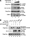
14-3-3 facilitates the Cby-CRM1 interaction but blocks the Cby-Importin-α interaction. (A) 14-3-3 promotes Cby binding to CRM1. Flag-CbyWT and increasing amounts of HA-14-3-3ζ were coexpressed in HEK293T cells, and cell lysates immunoprecipitated with anti-CRM1 antibody, followed by Western blot analysis with anti-Flag antibody. Immunoblotting of cell lysates with the indicated antibodies was performed to monitor protein levels. (B) 14-3-3 inhibits Cby binding to importin-α. Bacterially produced GST or GST-importin-α3 was incubated with cell lysates from HEK293T cells expressing Flag-tagged CbyWT or CbyS20A incapable of 14-3-3 binding and from those expressing HA-14-3-3ζ. The samples were then pulled down with glutathione beads, separated by SDS-PAGE, and immunoblotted with anti-Flag antibody.
Figure 7.
Figure 7.
Cby shuttling controls β-catenin localization and function. (A) CbyΔNLS2 sequesters β-catenin in the cytoplasm, whereas CbyL21A/L24A/L27A traps β-catenin in the nucleus. Cos7 cells were cotransfected with stabilized β-catenin-Myc and Flag-tagged CbyWT, CbyΔNLS2, or CbyL21A/L24A/L27A, and double-stained with anti-Cby (red) and anti-Myc (green) antibodies. Nuclei were visualized with DAPI. (B) Quantitative analysis of the results in A. The subcellular distribution of β-catenin-Myc was scored as described in the legend to Figure 1C. Error bars, means ± SD of three independent experiments. (C and D) Effects of Cby NLS mutants (C) or Cby NES mutants (D) on β-catenin–mediated transcriptional activation were evaluated by TopFlash assays. HEK293T cells were transfected with 10 ng of TopFlash luciferase reporter with or without 10 ng of an expression vector for stabilized β-catenin (β-catenin-Myc) and the indicated amounts of a Flag-tagged Cby expression plasmid. Luciferase activity was measured 24 h after transfection and normalized to Renilla luciferase activity used as an internal control. Transfections were carried out in triplicate, and the means ± SD are shown. The basal TopFlash value was set as 1. Western blot analysis with anti-Flag antibody showed that Cby proteins were expressed at similar levels (bottom panels). Note that, to compensate protein levels, higher amounts of DNA for CbyL21A, CbyL24A, CbyL27A, and CbyL21A/L24A/L27A were used for transfection.
Figure 8.
Figure 8.
Cby knockdown leads to nuclear accumulation of β-catenin. (A) Reduction of endogenous Cby protein levels by shRNA. Total cell lysates from HEK293 stable cell lines expressing control scrambled shRNA or Cby shRNA were immunoblotted with antibodies against Cby or GAPDH (loading control) as indicated. (B) β-Catenin accumulates in the nucleus in Cby-knockdown cells. Control or Cby shRNA-expressing HEK293 cells were transiently transfected with stabilized β-cateninS33Y-Flag and stained with anti-β-catenin antibody 17 h after transfection. Nuclei were labeled with DAPI. Similar results were obtained using anti-Flag antibody. (C) Quantitative analysis of the results in B. The subcellular distribution of β-catenin was scored as described in the legend to Figure 1C. Error bars, means ± SD of three independent experiments.

Similar articles

Cited by

References

    1. Aitken A. 14-3-3 proteins: a historic overview. Semin. Cancer Biol. 2006;16:162–172. - PubMed
    1. Barker N., Clevers H. Mining the Wnt pathway for cancer therapeutics. Nat. Rev. Drug Discov. 2006;5:997–1014. - PubMed
    1. Benzeno S., Diehl J. A. C-terminal sequences direct cyclin D1-CRM1 binding. J. Biol. Chem. 2004;279:56061–56066. - PubMed
    1. Bogerd H. P., Echarri A., Ross T. M., Cullen B. R. Inhibition of human immunodeficiency virus Rev and human T-cell leukemia virus Rex function, but not Mason-Pfizer monkey virus constitutive transport element activity, by a mutant human nucleoporin _targeted to Crm1. J. Virol. 1998;72:8627–8635. - PMC - PubMed
    1. Brunet A., Kanai F., Stehn J., Xu J., Sarbassova D., Frangioni J. V., Dalal S. N., DeCaprio J. A., Greenberg M. E., Yaffe M. B. 14-3-3 transits to the nucleus and participates in dynamic nucleocytoplasmic transport. J. Cell Biol. 2002;156:817–828. - PMC - PubMed

Publication types

MeSH terms

LinkOut - more resources

  NODES
INTERN 1
Note 4
twitter 2
Verify 1