Nanostructured polymer scaffolds for tissue engineering and regenerative medicine
- PMID: 20049793
- PMCID: PMC2800311
- DOI: 10.1002/wnan.26
Nanostructured polymer scaffolds for tissue engineering and regenerative medicine
Abstract
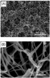
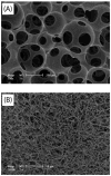
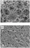
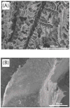
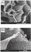
The structural features of tissue engineering scaffolds affect cell response and must be engineered to support cell adhesion, proliferation and differentiation. The scaffold acts as an interim synthetic extracellular matrix (ECM) that cells interact with prior to forming a new tissue. In this review, bone tissue engineering is used as the primary example for the sake of brevity. We focus on nanofibrous scaffolds and the incorporation of other components including other nanofeatures into the scaffold structure. Since the ECM is comprised in large part of collagen fibers, between 50 and 500 nm in diameter, well-designed nanofibrous scaffolds mimic this structure. Our group has developed a novel thermally induced phase separation (TIPS) process in which a solution of biodegradable polymer is cast into a porous scaffold, resulting in a nanofibrous pore-wall structure. These nanoscale fibers have a diameter (50-500 nm) comparable to those collagen fibers found in the ECM. This process can then be combined with a porogen leaching technique, also developed by our group, to engineer an interconnected pore structure that promotes cell migration and tissue ingrowth in three dimensions. To improve upon efforts to incorporate a ceramic component into polymer scaffolds by mixing, our group has also developed a technique where apatite crystals are grown onto biodegradable polymer scaffolds by soaking them in simulated body fluid (SBF). By changing the polymer used, the concentration of ions in the SBF and by varying the treatment time, the size and distribution of these crystals are varied. Work is currently being done to improve the distribution of these crystals throughout three-dimensional scaffolds and to create nanoscale apatite deposits that better mimic those found in the ECM. In both nanofibrous and composite scaffolds, cell adhesion, proliferation and differentiation improved when compared to control scaffolds. Additionally, composite scaffolds showed a decrease in incidence of apoptosis when compared to polymer control in bone tissue engineering. Nanoparticles have been integrated into the nanostructured scaffolds to deliver biologically active molecules such as growth and differentiation factors to regulate cell behavior for optimal tissue regeneration.
(c) 2009 John Wiley & Sons, Inc.
Figures

Similar articles
-
Biomimetic nanofibrous gelatin/apatite composite scaffolds for bone tissue engineering.Biomaterials. 2009 Apr;30(12):2252-8. doi: 10.1016/j.biomaterials.2008.12.068. Epub 2009 Jan 18. Biomaterials. 2009. PMID: 19152974 Free PMC article.
-
Phase separation, pore structure, and properties of nanofibrous gelatin scaffolds.Biomaterials. 2009 Sep;30(25):4094-103. doi: 10.1016/j.biomaterials.2009.04.024. Epub 2009 May 23. Biomaterials. 2009. PMID: 19481080 Free PMC article.
-
Macroporous and nanofibrous polymer scaffolds and polymer/bone-like apatite composite scaffolds generated by sugar spheres.J Biomed Mater Res A. 2006 Aug;78(2):306-15. doi: 10.1002/jbm.a.30704. J Biomed Mater Res A. 2006. PMID: 16637043
-
Nanostructured polymeric scaffolds for orthopaedic regenerative engineering.IEEE Trans Nanobioscience. 2012 Mar;11(1):3-14. doi: 10.1109/TNB.2011.2179554. Epub 2012 Jan 23. IEEE Trans Nanobioscience. 2012. PMID: 22275722 Review.
-
Design and fabrication of porous biodegradable scaffolds: a strategy for tissue engineering.J Biomater Sci Polym Ed. 2017 Nov;28(16):1797-1825. doi: 10.1080/09205063.2017.1354674. Epub 2017 Jul 24. J Biomater Sci Polym Ed. 2017. PMID: 28707508 Review.
Cited by
-
Bone Physiology, Biomaterial and the Effect of Mechanical/Physical Microenvironment on MSC Osteogenesis: A Tribute to Shu Chien's 80th Birthday.Cell Mol Bioeng. 2011 Dec;4(4):579-590. doi: 10.1007/s12195-011-0204-9. Cell Mol Bioeng. 2011. PMID: 25580165 Free PMC article.
-
Endothelial differentiation of human stem cells seeded onto electrospun polyhydroxybutyrate/polyhydroxybutyrate-co-hydroxyvalerate fiber mesh.PLoS One. 2012;7(4):e35422. doi: 10.1371/journal.pone.0035422. Epub 2012 Apr 16. PLoS One. 2012. PMID: 22523594 Free PMC article.
-
Anatase Incorporation to Bioactive Scaffolds Based on Salmon Gelatin and Its Effects on Muscle Cell Growth.Polymers (Basel). 2020 Aug 28;12(9):1943. doi: 10.3390/polym12091943. Polymers (Basel). 2020. PMID: 32872101 Free PMC article.
-
Conventional and Recent Trends of Scaffolds Fabrication: A Superior Mode for Tissue Engineering.Pharmaceutics. 2022 Jan 27;14(2):306. doi: 10.3390/pharmaceutics14020306. Pharmaceutics. 2022. PMID: 35214038 Free PMC article. Review.
-
Development of a PCL/gelatin/chitosan/β-TCP electrospun composite for guided bone regeneration.Prog Biomater. 2018 Sep;7(3):225-237. doi: 10.1007/s40204-018-0098-x. Epub 2018 Sep 21. Prog Biomater. 2018. PMID: 30242739 Free PMC article.
References
-
- Wang S, Zinderman C, Wise R, Braun M. Infections and human tissue transplants: review of FDA MedWatch reports 2001–2004. Cell Tissue Banking. 2007;8:211–219. - PubMed
-
- Laurencin CT, Khan Y. Bone graft substitute materials. eMedicine.com Inc; 2006. http://www.emedicine.com/orthoped/topic611.htm.
-
- Langer R, Vacanti JP. Tissue engineering. Science. 1993;260:920–926. - PubMed
-
- Chen VJ, Ma PX. Nano-fibrous poly(L-lactic acid) scaffolds with interconnected spherical macropores. Biomaterials. 2004;25(11):2065–2073. - PubMed
-
- Liu XH, Ma PX. Polymeric scaffolds for bone tissue engineering. Annals of Biomedical Engineering. 2004;32(3):477–486. - PubMed
Publication types
MeSH terms
Substances
Grants and funding
- T90 DK070071-05/DK/NIDDK NIH HHS/United States
- R01 DE017689/DE/NIDCR NIH HHS/United States
- R01 DE015384-05A2/DE/NIDCR NIH HHS/United States
- R01 DE017689-02/DE/NIDCR NIH HHS/United States
- P20GM069985/GM/NIGMS NIH HHS/United States
- P60 DK020572/DK/NIDDK NIH HHS/United States
- T32DE07057/DE/NIDCR NIH HHS/United States
- R01 DE017689-03/DE/NIDCR NIH HHS/United States
- DE015384/DE/NIDCR NIH HHS/United States
- T32 DE007057-32/DE/NIDCR NIH HHS/United States
- P20 GM069985/GM/NIGMS NIH HHS/United States
- T90 DK070071/DK/NIDDK NIH HHS/United States
- R21 GM075840-03/GM/NIGMS NIH HHS/United States
- T90DK070071/DK/NIDDK NIH HHS/United States
- R01 DE015384-04/DE/NIDCR NIH HHS/United States
- GM075849/GM/NIGMS NIH HHS/United States
- P30 DK092926/DK/NIDDK NIH HHS/United States
- DE017689/DE/NIDCR NIH HHS/United States
- R21 GM075840/GM/NIGMS NIH HHS/United States
- P60 DK020572-29S1/DK/NIDDK NIH HHS/United States
- P20 GM069985-04/GM/NIGMS NIH HHS/United States
- R21 GM075840-02/GM/NIGMS NIH HHS/United States
- R01 DE015384/DE/NIDCR NIH HHS/United States
- T32 DE007057/DE/NIDCR NIH HHS/United States
- P60DK020572-29S1/DK/NIDDK NIH HHS/United States
LinkOut - more resources
Full Text Sources
Other Literature Sources
Miscellaneous

