Generating a prion with bacterially expressed recombinant prion protein
- PMID: 20110469
- PMCID: PMC2893558
- DOI: 10.1126/science.1183748
Generating a prion with bacterially expressed recombinant prion protein
Abstract
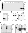
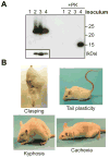
The prion hypothesis posits that a misfolded form of prion protein (PrP) is responsible for the infectivity of prion disease. Using recombinant murine PrP purified from Escherichia coli, we created a recombinant prion with the attributes of the pathogenic PrP isoform: aggregated, protease-resistant, and self-perpetuating. After intracerebral injection of the recombinant prion, wild-type mice developed neurological signs in approximately 130 days and reached the terminal stage of disease in approximately 150 days. Characterization of diseased mice revealed classic neuropathology of prion disease, the presence of protease-resistant PrP, and the capability of serially transmitting the disease; these findings confirmed that the mice succumbed to prion disease. Thus, as postulated by the prion hypothesis, the infectivity in mammalian prion disease results from an altered conformation of PrP.
Figures


Comment in
-
Biochemistry. What makes a prion infectious?Science. 2010 Feb 26;327(5969):1091-2. doi: 10.1126/science.1187790. Science. 2010. PMID: 20185716 No abstract available.
Similar articles
-
Conversion of bacterially expressed recombinant prion protein.Methods. 2011 Mar;53(3):208-13. doi: 10.1016/j.ymeth.2010.12.013. Epub 2010 Dec 19. Methods. 2011. PMID: 21176786 Free PMC article.
-
Synthetic mammalian prions.Science. 2004 Jul 30;305(5684):673-6. doi: 10.1126/science.1100195. Science. 2004. PMID: 15286374
-
Recombinant prion protein refolded with lipid and RNA has the biochemical hallmarks of a prion but lacks in vivo infectivity.PLoS One. 2013 Jul 30;8(7):e71081. doi: 10.1371/journal.pone.0071081. Print 2013. PLoS One. 2013. PMID: 23936256 Free PMC article.
-
The state of the prion.Nat Rev Microbiol. 2004 Nov;2(11):861-71. doi: 10.1038/nrmicro1025. Nat Rev Microbiol. 2004. PMID: 15494743 Review.
-
A general model of prion strains and their pathogenicity.Science. 2007 Nov 9;318(5852):930-6. doi: 10.1126/science.1138718. Science. 2007. PMID: 17991853 Review.
Cited by
-
Interactions between the conserved hydrophobic region of the prion protein and dodecylphosphocholine micelles.J Biol Chem. 2012 Jan 13;287(3):1915-22. doi: 10.1074/jbc.M111.279364. Epub 2011 Nov 29. J Biol Chem. 2012. PMID: 22128151 Free PMC article.
-
Genesis of mammalian prions: from non-infectious amyloid fibrils to a transmissible prion disease.PLoS Pathog. 2011 Dec;7(12):e1002419. doi: 10.1371/journal.ppat.1002419. Epub 2011 Dec 1. PLoS Pathog. 2011. PMID: 22144901 Free PMC article.
-
Aldehyde Production as a Calibrant of Ultrasonic Power Delivery During Protein Misfolding Cyclic Amplification.Protein J. 2020 Oct;39(5):501-508. doi: 10.1007/s10930-020-09920-1. Epub 2020 Oct 3. Protein J. 2020. PMID: 33011953
-
Glycoform-independent prion conversion by highly efficient, cell-based, protein misfolding cyclic amplification.Sci Rep. 2016 Jul 7;6:29116. doi: 10.1038/srep29116. Sci Rep. 2016. PMID: 27384922 Free PMC article.
-
Cofactor molecules: Essential partners for infectious prions.Prog Mol Biol Transl Sci. 2020;175:53-75. doi: 10.1016/bs.pmbts.2020.07.009. Epub 2020 Aug 24. Prog Mol Biol Transl Sci. 2020. PMID: 32958241 Free PMC article. Review.
References
Publication types
MeSH terms
Substances
Grants and funding
LinkOut - more resources
Full Text Sources
Other Literature Sources
Molecular Biology Databases
Research Materials

