. 2010 Jul;299(1):L59-72.
doi: 10.1152/ajplung.00380.2009. Epub 2010 Apr 9.

Chronic lung disease in preterm lambs: effect of daily vitamin A treatment on alveolarization

Affiliations

Chronic lung disease in preterm lambs: effect of daily vitamin A treatment on alveolarization

Kurt H Albertine et al. Am J Physiol Lung Cell Mol Physiol. 2010 Jul.

Abstract

Neonatal chronic lung disease is characterized by failed formation of alveoli and capillaries, and excessive deposition of matrix elastin, which are linked to lengthy mechanical ventilation (MV) with O(2)-rich gas. Vitamin A supplementation has improved respiratory outcome of premature infants, but there is little information about the structural and molecular manifestations in the lung that occur with vitamin A treatment. We hypothesized that vitamin A supplementation during prolonged MV, without confounding by antenatal steroid treatment, would improve alveolar secondary septation, decrease thickness of the mesenchymal tissue cores between distal air space walls, and increase alveolar capillary growth. We further hypothesized that these structural advancements would be associated with modulated expression of tropoelastin and deposition of matrix elastin, phosphorylated Smad2 (pSmad2), cleaved caspase 3, proliferating cell nuclear antigen (PCNA), VEGF, VEGF-R2, and midkine in the parenchyma of the immature lung. Eight preterm lambs (125 days' gestation, term approximately 150 days) were managed by MV for 3 wk: four were treated with daily intramuscular Aquasol A (vitamin A), 5,000 IU/kg, starting at birth; four received vehicle alone. Postmortem lung assays included quantitative RT-PCR and in situ hybridization, immunoblot and immunohistochemistry, and morphometry and stereology. Daily vitamin A supplementation increased alveolar secondary septation, decreased thickness of the mesenchymal tissue cores between the distal air space walls, and increased alveolar capillary growth. Associated molecular changes were less tropoelastin mRNA expression, matrix elastin deposition, pSmad2, and PCNA protein localization in the mesenchymal tissue core of the distal air space walls. On the other hand, mRNA expression and protein abundance of VEGF, VEGF-R2, midkine, and cleaved caspase 3 were increased. We conclude that vitamin A treatment partially improves lung development in chronically ventilated preterm neonates by modulating expression of tropoelastin, deposition of elastin, and expression of vascular growth factors.


Figures

Fig. 1.
Fig. 1.
Plasma retinol concentration. Daily vitamin A treatment (+ Vitamin A) of preterm lambs during 3 wk of mechanical ventilation (MV) increased plasma retinol concentration compared with vehicle controls (*P < 0.05; means ± SD; n = 4/group). Plasma retinol concentration in the vitamin A-treated preterm lambs was equivalent to that in lambs that were born at term gestation (Term). Term lambs served as the gestation age-matched reference for the 3-wk ventilation studies. Plasma retinol concentration in the vehicle controls was low at birth (d0) and remained low at the conclusion of the 3-wk study. That group's plasma retinol concentration is equal to that of fetal lambs that were delivered at 125 days of gestation (f125). Those fetal lambs served as the gestation age-matched reference at the time the preterm lambs were delivered. †Significant difference compared with the paired plasma concentration for the same group at d0 of life, P < 0.05. ‡Significant difference compared with the f125 reference group, P < 0.05.
Fig. 2.
Fig. 2.
Terminal respiratory unit (TRU) architecture. A preterm lamb treated daily with vitamin A (+ Vitamin A; B) during 3 wk of MV has TRU architecture that is heterogeneous. The proximal parts (respiratory bronchioles and initial segments of alveolar ducts) have alveolar simplification (*). That is, the air spaces are distended, alveolar secondary septa are infrequent, and those that are present are stunted, and the air space walls are thick and cellular. The distal parts of the same TRU have air spaces that are small, alveolar secondary septa that are numerous, long, and thin (arrows), and air space walls that are thin and less cellular (arrowhead). The TRU in the vehicle control (− Vitamin A; A) has uniformly distended air spaces, few alveolar secondary septa, and those that are present are short and thick (arrows), and distal air space walls are thick and cellular (arrowhead). A and B are the same magnification. Quantitative histology (C–E) showed that radial alveolar count (C) and volume density of alveolar secondary septa (Vv2′ septa; D) are greater in vitamin A-supplemented preterm lambs compared with vehicle controls (*P < 0.05; means ± SD; n = 4/group). However, radial alveolar count and Vv2′ septa in the vitamin A-treated preterm lambs is less than in lambs born at term gestation (Term; ◊P < 0.05). Term lambs served as the gestation age-matched reference for the 3-wk ventilation studies. Mean face length (E), on the other hand, is shorter in the vitamin A-treated preterm lambs than in the vehicle controls (*P < 0.05; means ± SD; n = 4/group); however, mean face length is longer than in lambs born at term gestation (Term; ◊P < 0.05). All 3 structural indices of alveolar formation in the vehicle controls are the same as in f125 reference group. ‡Significant difference compared with the f125 reference group, P < 0.05. ◊Significant difference compared with both preterm groups, P < 0.05.
Fig. 3.
Fig. 3.
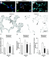
Elastin expression and deposition. A preterm lamb treated daily with vitamin A (+ Vitamin A; B) during 3 wk of MV has less tropoelastin mRNA localization in the lung parenchyma by in situ hybridization (green in B) and matrix elastin deposition revealed by Hart's stain (arrows in E) than a vehicle control (− Vitamin A; A and D, respectively). C is the sense control for in situ hybridization (blue fluorescence is DAPI-stained nuclei). No green immunofluorescence is visible, indicating specificity of the antisense probe used for tropoelastin. A–C are the same magnification, as are D and E. Daily vitamin A treatment quantitatively stabilized tropoelastin mRNA expression (F), desmosine content (G), and parenchymal elastic fiber volume density (H) compared with vehicle controls (− Vitamin A). However, expression and protein abundance in the vitamin A-treated preterm lambs is greater than in lambs born at term gestation (Term; ◊P < 0.05). Term lambs served as the gestation age-matched reference for the 3-wk ventilation studies. ‡Significant difference compared with the f125 reference group, P < 0.05. ◊Significant difference compared with both preterm groups, P < 0.05.
Fig. 4.
Fig. 4.
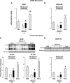
VEGF and VEGF-R2 expression. Daily vitamin A treatment (+ Vitamin A) of preterm lambs during 3 wk of MV increased VEGF-A and VEGF-R2 mRNA expression (A and B, respectively) and protein abundance (C and D, respectively) compared with vehicle (− Vitamin A) preterm controls (*P < 0.05; means ± SD; n = 4/group). However, mRNA expression and protein abundance in the vitamin A-treated preterm lambs is less than in lambs born at term gestation (Term; ◊P < 0.05). Term lambs served as the gestation age-matched reference for the 3-wk ventilation studies. VEGF and VEGF-R2 mRNA expression and protein abundance in the vehicle controls are the lowest among the groups. VEGF-A and VEGF-R2 mRNA expression is normalized for GAPDH mRNA expression, whereas abundance of the respective proteins is normalized for endogenous protein revealed by MemCode reversible protein stain. ‡Significant difference compared with the f125 reference group, P < 0.05. ◊Significant difference compared with both preterm groups, P < 0.05.
Fig. 5.
Fig. 5.
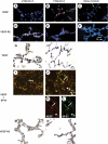
Localization of VEGF and VEGF-R2. A preterm lamb treated daily with vitamin A (+ Vitamin A) during 3 wk of MV has more VEGF-A and VEGF-R2 mRNA expression visible (red fluorescence in B and E, respectively) compared with a vehicle control (− Vitamin A; A and D, respectively). C and F are the sense controls for in situ hybridization (blue fluorescence is DAPI-stained nuclei). No red immunofluorescence is visible, indicating specificity of the antisense probe used for VEGF-A and VEGF-R2, respectively. A–F are the same magnification. In lung tissue from the same preterm lamb treated daily with vitamin A, more VEGF and VEGF-R2 protein localization is visible (brown immunostain in H and N, respectively) compared with the vehicle control (G and M, respectively). G, H, M, and N are the same magnification. VEGF protein that is immunolocalized in domed epithelial cells in G and H (arrows) is specifically localized in alveolar type II epithelial cells (yellow fluorescence in I and J) because VEGF protein (red fluorescence in K) colocalized with SP-B protein (green fluorescence in L). I and J are the same magnification. VEGF-R2 protein is immunolocalized in capillary endothelial cells (arrowhead in N) and other cells such as vascular smooth muscle cells and epithelial cells (arrows of M and N).
Fig. 6.
Fig. 6.
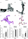
Alveolar secondary septal ultrastructure and capillary localization. A preterm lamb treated daily with vitamin A (+ Vitamin A; B) during 3 wk of MV has an alveolar secondary septum (Sec Septum) that is longer, thinner, and has capillaries (C) along its length compared with the vehicle control (A; shown at the same magnification as B). Alv Wall, alveolar wall; E, elastin; M, mesenchymal cell. Immunohistochemical localization of PECAM-1 protein highlights endothelial cells with brown immunostain (arrows in C and D; shown at the same magnification). Daily vitamin A treatment quantitatively increased capillary surface density (E), referenced to equal epithelial cells surface density (F), and extra-alveolar microvessel number/100 parenchymal points (>20 but <100 μm in diameter; G) compared with vehicle controls (− Vitamin A; *P < 0.05; means ± SD; n = 4/group). However, capillary surface density and microvessel number in the vitamin A-treated preterm lambs is less than in lambs born at term gestation (Term; ◊P < 0.05). Term lambs served as the gestation age-matched reference for the 3-wk ventilation studies. ‡Significant difference compared with the f125 reference group, P < 0.05. ◊Significant difference compared with both preterm groups, P < 0.05.
Fig. 7.
Fig. 7.
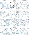
Localization of midkine, cleaved caspase 3, PCNA, and pSmad2. A preterm lamb treated daily with vitamin A (+ Vitamin A) during 3 wk of MV has more midkine and cleaved caspase 3 protein localization visible among mesenchymal cells (brown stain in B and E, respectively) compared with a vehicle control (− Vitamin A; A and D, respectively). Conversely, lung tissue sections from the vitamin A-treated preterm lamb have less immunostain for PCNA and pSmad2 among mesenchymal cells (brown stain in H and K, respectively) compared with the vehicle control (G and J, respectively). Immunostained cells in the mesenchyme of the distal air space (DAS) walls are labeled with small, thin arrows. The bold arrow in the panels with an inset image identifies the region illustrated at greater magnification in the corresponding inset image. All panels are the same magnification. All inset images are the same magnification (twice the magnification of the panels). The column of images on the right shows immunohistochemical staining results for a term newborn lamb, as a gestation age-matched reference for the preterm lambs. Immunolocalization patterns for midkine (C), cleaved caspase 3 (F), PCNA (I), and pSmad2 (L) are most similar to the preterm lamb treated daily with vitamin A during 3 wk of MV.

Similar articles

Cited by

References

    1. Abman SH. Bronchopulmonary dysplasia: “a vascular hypothesis.” Am J Respir Crit Care Med 164: 1755–1756, 2001 - PubMed
    1. Albertine KH, Dahl M, Plopper CG, Hyde DM. Stereological approaches to quantify alveolar formation in the lung. FASEB J 17: A775, 2003
    1. Albertine KH, Jones GP, Starcher BC, Bohnsack JF, Davis PL, Cho SC, Carlton DP, Bland RD. Chronic lung injury in preterm lambs: disordered respiratory tract development. Am J Respir Crit Care Med 159: 945–958, 1999 - PubMed
    1. Albertine KH, Pysher TJ. Impaired lung growth after injury in premature lung. In: Fetal and Neonatal Physiology ( 3rd ed.), edited by Polin RA, Fox WW, Abman SH. New York: Saunders, 2004, p. 942–949
    1. Albertine KH, Soulier MF, Wang Z, Ishizaka A, Hashimoto S, Zimmerman GA, Matthay MA, Ware LB. Fas and fas ligand are up-regulated in pulmonary edema fluid and lung tissue of patients with acute lung injury and the acute respiratory distress syndrome. Am J Pathol 161: 1783–1796, 2002 - PMC - PubMed

Publication types

MeSH terms

  NODES
twitter 2