Propensity to high-fat diet-induced obesity in rats is associated with changes in the gut microbiota and gut inflammation
- PMID: 20508158
- PMCID: PMC2928532
- DOI: 10.1152/ajpgi.00098.2010
Propensity to high-fat diet-induced obesity in rats is associated with changes in the gut microbiota and gut inflammation
Abstract
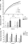
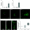
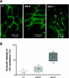
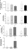
Consumption of diets high in fat and calories leads to hyperphagia and obesity, which is associated with chronic "low-grade" systemic inflammation. Ingestion of a high-fat diet alters the gut microbiota, pointing to a possible role in the development of obesity. The present study used Sprague-Dawley rats that, when fed a high-fat diet, exhibit either an obesity-prone (DIO-P) or obesity-resistant (DIO-R) phenotype, to determine whether changes in gut epithelial function and microbiota are diet or obese associated. Food intake and body weight were monitored daily in rats maintained on either low- or high-fat diets. After 8 or 12 wk, tissue was removed to determine adiposity and gut epithelial function and to analyze the gut microbiota using PCR. DIO-P but not DIO-R rats exhibit an increase in toll-like receptor (TLR4) activation associated with ileal inflammation and a decrease in intestinal alkaline phosphatase, a luminal enzyme that detoxifies lipopolysaccharide (LPS). Intestinal permeability and plasma LPS were increased together with phosphorylation of myosin light chain and localization of occludin in the cytoplasm of epithelial cells. Measurement of bacterial 16S rRNA showed a decrease in total bacterial density and an increase in the relative proportion of Bacteroidales and Clostridiales orders in high-fat-fed rats regardless of phenotype; an increase in Enterobacteriales was seen in the microbiota of DIO-P rats only. Consumption of a high-fat diet induces changes in the gut microbiota, but it is the development of inflammation that is associated with the appearance of hyperphagia and an obese phenotype.
Figures


Similar articles
-
Diet-driven microbiota dysbiosis is associated with vagal remodeling and obesity.Physiol Behav. 2017 May 1;173:305-317. doi: 10.1016/j.physbeh.2017.02.027. Epub 2017 Feb 27. Physiol Behav. 2017. PMID: 28249783 Free PMC article.
-
Apple-Derived Pectin Modulates Gut Microbiota, Improves Gut Barrier Function, and Attenuates Metabolic Endotoxemia in Rats with Diet-Induced Obesity.Nutrients. 2016 Feb 29;8(3):126. doi: 10.3390/nu8030126. Nutrients. 2016. PMID: 26938554 Free PMC article.
-
Alterations to the microbiota-colon-brain axis in high-fat-diet-induced obese mice compared to diet-resistant mice.J Nutr Biochem. 2019 Mar;65:54-65. doi: 10.1016/j.jnutbio.2018.08.016. Epub 2018 Sep 1. J Nutr Biochem. 2019. PMID: 30623851
-
Translational research into gut microbiota: new horizons in obesity treatment.Arq Bras Endocrinol Metabol. 2009 Mar;53(2):139-44. doi: 10.1590/s0004-27302009000200004. Arq Bras Endocrinol Metabol. 2009. Retraction in: Arq Bras Endocrinol Metabol. 2013 Dec;57(9):753. doi: 10.1590/s0004-27302013000900014 PMID: 19466205 Retracted. Review.
-
Impact of dietary fat on gut microbiota and low-grade systemic inflammation: mechanisms and clinical implications on obesity.Int J Food Sci Nutr. 2018 Mar;69(2):125-143. doi: 10.1080/09637486.2017.1343286. Epub 2017 Jul 4. Int J Food Sci Nutr. 2018. PMID: 28675945 Review.
Cited by
-
Effects of dietary components on intestinal permeability in health and disease.Am J Physiol Gastrointest Liver Physiol. 2020 Nov 1;319(5):G589-G608. doi: 10.1152/ajpgi.00245.2020. Epub 2020 Sep 9. Am J Physiol Gastrointest Liver Physiol. 2020. PMID: 32902315 Free PMC article. Review.
-
Obesity and adipose tissue impact on T-cell response and cancer immune checkpoint blockade therapy.Immunother Adv. 2022 Jun 24;2(1):ltac015. doi: 10.1093/immadv/ltac015. eCollection 2022. Immunother Adv. 2022. PMID: 36033972 Free PMC article. Review.
-
The Effect of High-Fat Diet-Induced Pathophysiological Changes in the Gut on Obesity: What Should be the Ideal Treatment?Clin Transl Gastroenterol. 2013 Jul 11;4(7):e39. doi: 10.1038/ctg.2013.11. Clin Transl Gastroenterol. 2013. PMID: 23842483 Free PMC article.
-
High fat diet causes depletion of intestinal eosinophils associated with intestinal permeability.PLoS One. 2015 Apr 2;10(4):e0122195. doi: 10.1371/journal.pone.0122195. eCollection 2015. PLoS One. 2015. PMID: 25837594 Free PMC article.
-
Composition of dietary fat source shapes gut microbiota architecture and alters host inflammatory mediators in mouse adipose tissue.JPEN J Parenter Enteral Nutr. 2013 Nov;37(6):746-54. doi: 10.1177/0148607113486931. Epub 2013 May 2. JPEN J Parenter Enteral Nutr. 2013. PMID: 23639897 Free PMC article.
References
-
- Alpers DH, Zhang Y, Ahnen DJ. Synthesis and parallel secretion of rat intestinal alkaline phosphatase and a surfactant-like particle protein. Am J Physiol Endocrinol Metab 268: E1205–E1214, 1995 - PubMed
-
- Amar J, Burcelin R, Ruidavets JB, Cani PD, Fauvel J, Alessi MC, Chamontin B, Ferrieres J. Energy intake is associated with endotoxemia in apparently healthy men. Am J Clin Nutr 87: 1219–1223, 2008 - PubMed
-
- Backhed F. Changes in intestinal microflora in obesity: cause or consequence? J Pediatr Gastroenterol Nutr 48, Suppl 2: S56–S57, 2009 - PubMed
Publication types
MeSH terms
Substances
Grants and funding
LinkOut - more resources
Full Text Sources
Other Literature Sources
Medical
Miscellaneous

