A subpopulation of CD163-positive macrophages is classically activated in psoriasis
- PMID: 20555352
- PMCID: PMC2939947
- DOI: 10.1038/jid.2010.165
A subpopulation of CD163-positive macrophages is classically activated in psoriasis
Abstract
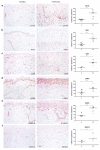
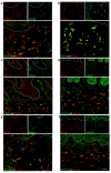
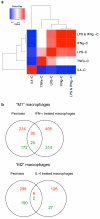
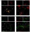
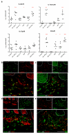
Macrophages are important cells of the innate immune system, and their study is essential to gain greater understanding of the inflammatory nature of psoriasis. We used immunohistochemistry and double-label immunofluorescence to characterize CD163(+) macrophages in psoriasis. Dermal macrophages were increased in psoriasis compared with normal skin and were identified by CD163, RFD7, CD68, lysosomal-associated membrane protein 2 (LAMP2), stabilin-1, and macrophage receptor with collagenous structure (MARCO). CD163(+) macrophages expressed C-lectins CD206/macrophage mannose receptor and CD209/DC-SIGN, as well as costimulatory molecules CD86 and CD40. They did not express mature dendritic cell (DC) markers CD208/DC-lysosomal-associated membrane glycoprotein, CD205/DEC205, or CD83. Microarray analysis of in vitro-derived macrophages treated with IFN-γ showed that many of the genes upregulated in macrophages were found in psoriasis, including STAT1, CXCL9, Mx1, and HLA-DR. CD163(+) macrophages produced inflammatory molecules IL-23p19 and IL-12/23p40 as well as tumor necrosis factor (TNF) and inducible nitric oxide synthase (iNOS). These data show that CD163 is a superior marker of macrophages, and identifies a subpopulation of "classically activated" macrophages in psoriasis. We conclude that macrophages are likely to contribute to the pathogenic inflammation in psoriasis, a prototypical T helper 1 (Th1) and Th17 disease, by releasing key inflammatory products.
Figures
Similar articles
-
Association of the numbers of CD163(+) cells in lesional skin and serum levels of soluble CD163 with disease progression of cutaneous T cell lymphoma.J Dermatol Sci. 2012 Oct;68(1):45-51. doi: 10.1016/j.jdermsci.2012.07.007. Epub 2012 Jul 27. J Dermatol Sci. 2012. PMID: 22884782
-
GM-CSF Expression and Macrophage Polarization in Joints of Undifferentiated Arthritis Patients Evolving to Rheumatoid Arthritis or Psoriatic Arthritis.Front Immunol. 2021 Feb 17;11:613975. doi: 10.3389/fimmu.2020.613975. eCollection 2020. Front Immunol. 2021. PMID: 33679701 Free PMC article.
-
Tumor-associated macrophages in oral premalignant lesions coexpress CD163 and STAT1 in a Th1-dominated microenvironment.BMC Cancer. 2015 Aug 5;15:573. doi: 10.1186/s12885-015-1587-0. BMC Cancer. 2015. PMID: 26242181 Free PMC article.
-
Macrophage Activation Markers, CD163 and CD206, in Acute-on-Chronic Liver Failure.Cells. 2020 May 9;9(5):1175. doi: 10.3390/cells9051175. Cells. 2020. PMID: 32397365 Free PMC article. Review.
-
The macrophage scavenger receptor CD163.Immunobiology. 2005;210(2-4):153-60. doi: 10.1016/j.imbio.2005.05.010. Immunobiology. 2005. PMID: 16164022 Review.
Cited by
-
Second-Strand Synthesis-Based Massively Parallel scRNA-Seq Reveals Cellular States and Molecular Features of Human Inflammatory Skin Pathologies.Immunity. 2020 Oct 13;53(4):878-894.e7. doi: 10.1016/j.immuni.2020.09.015. Immunity. 2020. PMID: 33053333 Free PMC article.
-
Omics-Driven Biomarkers of Psoriasis: Recent Insights, Current Challenges, and Future Prospects.Clin Cosmet Investig Dermatol. 2020 Aug 25;13:611-625. doi: 10.2147/CCID.S227896. eCollection 2020. Clin Cosmet Investig Dermatol. 2020. PMID: 32922059 Free PMC article. Review.
-
Using Micro- and Macro-Level Network Metrics Unveils Top Communicative Gene Modules in Psoriasis.Genes (Basel). 2020 Aug 10;11(8):914. doi: 10.3390/genes11080914. Genes (Basel). 2020. PMID: 32785106 Free PMC article.
-
Macrophage polarization reflects T cell composition of tumor microenvironment in pediatric classical Hodgkin lymphoma and has impact on survival.PLoS One. 2015 May 15;10(5):e0124531. doi: 10.1371/journal.pone.0124531. eCollection 2015. PLoS One. 2015. PMID: 25978381 Free PMC article.
-
Doppler ultrasound-based noninvasive biomarkers in hidradenitis suppurativa: evaluation of analytical and clinical validity.Br J Dermatol. 2021 Apr;184(4):688-696. doi: 10.1111/bjd.19343. Epub 2020 Sep 6. Br J Dermatol. 2021. PMID: 32602132 Free PMC article.
References
-
- Arredouani MS, Palecanda A, Koziel H, Huang YC, Imrich A, Sulahian TH, et al. MARCO is the major binding receptor for unopsonized particles and bacteria on human alveolar macrophages. J Immunol. 2005;175:6058–6064. - PubMed
-
- Bryant P, Ploegh H. Class II MHC peptide loading by the professionals. Curr Opin Immunol. 2004;16:96–102. - PubMed
-
- Djemadji-Oudjiel N, Goerdt S, Kodelja V, Schmuth M, Orfanos CE. Immunohistochemical identification of type II alternatively activated dendritic macrophages (RM 3/1+3, MS-1+/−, 25F9-) in psoriatic dermis. Arch Dermatol Res. 1996;288:757–764. - PubMed
Publication types
MeSH terms
Substances
Grants and funding
LinkOut - more resources
Full Text Sources
Other Literature Sources
Medical
Research Materials
Miscellaneous

