. 2011 Feb;18(2):350-61.
doi: 10.1038/cdd.2010.104. Epub 2010 Aug 27.

Sphingosine-1-phosphate phosphohydrolase-1 regulates ER stress-induced autophagy

Affiliations

Sphingosine-1-phosphate phosphohydrolase-1 regulates ER stress-induced autophagy

S Lépine et al. Cell Death Differ. 2011 Feb.

Abstract

The sphingolipid metabolites ceramide and sphingosine-1-phosphate (S1P) have recently been implicated in autophagy. In this study, we report that depletion of sphingosine-1-phosphate phosphohydrolase-1 (SPP1), an endoplasmic reticulum (ER)-resident enzyme that specifically dephosphorylates S1P, induced autophagy. Although the mammalian _target of rapamycin and class III phosphoinositide 3-kinase/Beclin-1 pathways were not involved and this autophagy was p53 independent, C/EBP homologous protein, BiP, and phospho-eucaryotic translation initiation factor-2α, and cleavage of procaspases 2 and 4, downstream _targets of ER stress, were increased after SPP1 depletion. Autophagy was suppressed by depletion of protein kinase regulated by RNA-like ER kinase (PERK), inositol-requiring transmembrane kinase/endonuclease-1α, or activating transcription factor 6, three sensors of the unfolded protein response (UPR) to ER stress. Autophagy triggered by downregulation of SPP1 did not lead to apoptosis but rather stimulated, in a PERK dependent manner, the survival signal Akt, whose inhibition then sensitized cells to apoptosis. Although depletion of SPP1 increased intracellular levels of S1P and its secretion, activation of cell surface S1P receptors did not induce autophagy. Nevertheless, increases in intracellular pools of S1P, but not dihydro-S1P, induced autophagy and ER stress. Thus, SPP1, by regulating intracellular S1P homeostasis, can control the UPR and ER stress-induced autophagy.


Figures
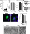
Figure 1
Figure 1
Depletion of SPP1 induces autophagy in MCF7 cells. MCF7 cells were transfected with control scrambled siRNA or ON-_targetplus SMARTpool siRNA _targeted to SPP1 (siSPP1) or with individual siRNAs _targeted to two distinct SPP1 sequences (siSPP1#1, siSPP1#2), as indicated. (a) SPP1, SPP2, and S1P lyase mRNA levels were determined by quantitative real-time PCR and normalized to glyceraldehyde-3-phosphate dehydrogenase (GAPDH) mRNA. (b, c) Lysates from MCF7 cells transfected with siControl or siSPP1 were analyzed by immunoblotting with anti-SPP1 antibody (b), or with anti-LC3 antibody (c) and re-probed with anti-tubulin antibody to insure equal loading and transfer. Numbers indicate fold changes determined by densitometry. (d, e) Twenty-four hours after siRNA transfections as in (a), cells were transfected with EGFP-LC3. After culturing for an additional 24 h in complete medium, cells were examined by confocal fluorescence microscopy. (d) Representative images are shown. Scale bars, 10 μm. (e) Percentage of cells showing GFP-LC3 fluorescence in puncta was quantified. Mock, untransfected cells. Data are means±S.D. from three independent experiments. At least 100 cells were analyzed for each. (f) MCF7 cells transfected with siControl or siSPP1 were examined by transmission electron microscopy. Representative micrographs are shown and atypical autophagosome are boxed and shown at higher magnification. Scale bars, 0.5 μm. *P<0.01 compared with siControl
Figure 2
Figure 2
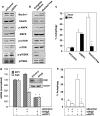
Downregulation of SPP1 does not activate the mTOR pathway. (a, c) MCF7 cells were transfected with siControl or siSPP1 or were cultured in nutrient-free EBSS or in complete medium, as indicated. (a, b) Equal amounts of cell lysates were resolved by SDS-PAGE and immunoblotted with the indicated antibodies. Blots were re-probed for total mTOR, AMPK, p70S6K, and tubulin to insure equal loading and transfer. (c) Cells treated as indicated in (a) were transfected with EGFP-LC3. After culturing for 24 h in complete medium or in nutrient-free medium where indicated, in the absence or presence of 10 mM 3MA, autophagy was quantified by confocal microscopy. Data are means±S.D. of three independent experiments. *P<0.01 compared with untreated. (d, e) MCF7 cells were transfected with control siRNA, siSPP1, or siAtg5, as indicated. (d) SPP1 and Atg5 mRNA levels were determined by quantitative real-time PCR and normalized to glyceraldehyde-3-phosphate dehydrogenase (GAPDH) mRNA. Insert: Equal amounts of cell lysates were immunoblotted with anti-Atg5 and re-probed for tubulin to insure equal loading and transfer. (e) Autophagy was determined in duplicate cultures transfected with EGFP-LC3. *P<0.01 compared with siControl
Figure 3
Figure 3
Downregulation of SPP1 induces ER stress that leads to autophagy. (a, b) Equal amounts of lysates from MCF7 cells transfected with siControl or siSPP1 were resolved by SDS-PAGE and immunoblotted with the indicated antibodies. Blots were re-probed for total eIF2α or tubulin to insure equal loading and transfer. (c) Duplicate cultures were lysed and caspase 2 and caspase 4 activities were determined by fluorescence and luminescence, respectively. Data are means±S.D. of three independent experiments each carried out in triplicate. (d) MCF7 cells were transfected with siControl or siRNAs _targeted to IRE1α or ATF6, and downregulation efficiency was examined by immunoblotting. (e) MCF7 cells were also co-transfected with vector or dominant-negative PERK (dnPERK) and inhibition of PERK activity confirmed by immunoblotting with p-eIF2α antibody. (f) MCF7 cells were co-transfected with siControl, siSPP1, siIRE1α, siATF6, or dnPERK and EGFP-LC3, as indicated. Autophagy was quantified by confocal microscopy. Values are the means±S.D. of three independent experiments. (gi) Downregulation of SPP1 does not trigger apoptosis or induce cell death. (g) MCF7 cells were transfected with siControl or siSPP1, cultured for the indicated times, and nuclei were stained with Hoechst 33342. Apoptosis was determined by scoring the percentage of cells displaying fragmented, condensed nuclei indicative of apoptosis. At least three fields were analyzed, scoring a minimum of 300 cells. Data are the means±S.D. of three independent experiments. Insert: Equal amounts of lysates from duplicate 1-day cultures were resolved by SDS-PAGE and immunoblotted with anti-PARP antibody. Blots were re-probed with anti-tubulin to insure equal loading and transfer. (h, i) MCF7 cells were transfected with siControl siRNA, siSPP1, or individual siRNAs _targeted to Atg5, ATF6, or IRE1α, or transfected with dnPERK. Apoptosis was quantified by Hoechst 33342 staining (h) and viability was quantified by Trypan blue exclusion (i). Data are means±S.D. of three independent experiments. *P<0.01 compared with siControl
Figure 4
Figure 4
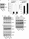
SPP1-mediated autophagy is p53-independent. (a) MCF7 cells were transfected with control siRNA or with siSPP1. Equal amounts of lysates were resolved by SDS-PAGE and immunoblotted with anti-p53 antibody. Blots were re-probed for tubulin to insure equal loading and transfer. (bf) Wild-type (WT) or p53−/− HCT116 cells were transfected with control siRNA or siSPP1. (b) SPP1 mRNA was determined by quantitative real-time PCR and normalized to GAPDH mRNA. (c) Twenty-four hours after siRNA transfections, as in (b), cells were transfected with EGFP-LC3 and autophagy was determined by confocal microscopy. *P<0.01 compared with siControl. (d) Autophagy was determined by western blotting with anti-LC3 antibody and with anti-tubulin as a loading control. Numbers indicate fold changes determined by densitometry. (e, f) Equal amounts of cell lysates were resolved by SDS-PAGE and immunoblotted with the indicated antibodies. Blots were re-probed for tubulin or total mTOR to insure equal loading and transfer
Figure 5
Figure 5
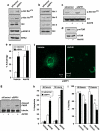
Downregulation of SPP1 stimulates Akt, which suppresses apoptosis. (a, b) MCF7 cells were transfected with siControl or siSPP1. (c, d) Cells were cultured for 24 h in the absence or presence of AktVIII (5 μM) as indicated. (ad) Equal amounts of cell lysates were resolved by SDS-PAGE and immunoblotted with the indicated antibodies. (e, f) MCF7 cells transfected with siControl or siSPP1 were transfected with EGFP-LC3 and cultured in the absence or presence of AktVIII (5 μM). Autophagy was determined by confocal microscopy. (f) Representative images are shown. Scale bars, 10 μm. Data are means±S.D. of three independent experiments. *P<0.05 compared with siControl. (gi) MCF7 cells transfected with siControl or siSPP1 were cultured in the absence or presence of AktVIII (5 μM) for 48 or 72 h. (g) Equal amounts of lysates were resolved by SDS-PAGE and immunoblotted with anti-PARP antibody. Apoptosis (h) and viability (i) were quantified after 48 and 72 h in duplicate cultures by Hoechst 33342 and Trypan blue exclusion staining, respectively. *P<0.05 compared with siControl, **P<0.05 compared with vehicle
Figure 6
Figure 6
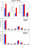
Effect of SPP1 depletion on S1P and sphingolipids. (ac) MCF7 cells were transfected with siControl or siSPP1. Lipids were extracted and S1P (a), dihydro-S1P (b), sphingosine and dihydrosphingosine (c), ceramide species (d), and dihydroceramide species (e) analyzed by LC-MS/MS. Numbers indicate chain length followed by the number of double bonds in the fatty acid. Data are averages of triplicate transfectants and are expressed as picomol lipid/mg of protein. *P<0.01 compared with siControl. Similar results were found in three additional experiments
Figure 7
Figure 7
SPP1 depletion triggers autophagy and activates Akt by intracellular S1P independently of its receptors. (a) MCF7 cells transfected with pEGFP-LC3 were treated with 100 nM S1P or dihydroS1P (DHS1P) for 24 h, and autophagy was quantified by confocal microscopy. Data are means±S.D. of three independent experiments. (b) Cells were treated with 100 nM S1P or DHS1P for 10 min. (c) Cells were pretreated without or with 10 μM VPC23019 or 1 μM JTE-013 for 30 min, and then stimulated with 100 nM S1P for 10 min. (b, c) Equal amounts of lysates were analyzed by immunoblotting with anti-phospho ERK1/2. Blots were re-probed for ERK to insure equal loading and transfer. (d) Cells transfected with siControl or siSPP1 were transfected 24 h later with EGFP-LC3, cultured in the absence or presence of 10 μM VPC23019 or 1 μM JTE-013, and autophagy quantified by confocal microscopy. Values reported are means±S.D. of three independent experiments. (e) MCF7 cells transfected with siControl or siSPP1 or naïve MCF7 cells were treated with vehicle, S1P (100 nM), or EGF (10 ng/ml) for 10 min. Proteins were immunoblotted with anti-phospho-Akt (Ser473) and re-probed with anti-Akt antibody. (f) MCF7 cells were treated with 10 μM S1P or dihydroS1P (DHS1P) for 2 h and sphingolipids analyzed by LC-ESI-MS/MS. (g) MCF7 cells transfected with pEGFP-LC3 were treated with 10 μM S1P or DHS1P for 24 h and autophagy quantified by confocal microscopy. (h) Equal amounts of lysates from MCF7 cells treated without or with 10 μM S1P were immunoblotted with anti-LC3 or anti-p-eIF2α antibodies. Blots were re-probed for tubulin to insure equal loading and transfer. (i) MCF7 cells transfected with siControl or siSPP1 were transfected with vector or dnPERK. (j) MCF7 cells transfected with siControl or siSPP1 were treated without or with LY294004 (5 μM). Equal amounts of lysates were immunoblotted with anti-phospho-Akt (Ser473) and re-probed with anti-Akt antibody. (k) Wild-type (PERK+/+) and PERK null (PERK−/−) MEFs were transfected with siControl or siSPP1. Equal amounts of lysates were immunoblotted with the indicated antibodies. *P<0.01 compared with vehicle
Figure 8
Figure 8
Model of involvement of SPP1 in ER stress and UPR and connection to autophagy. Depletion of SPP1 and increased intracellular S1P triggers ER stress and the UPR by activation of ATF6, IRE1α, and PERK, resulting in autophagy. PERK signaling also activates Akt, which protect cells from apoptosis. Other known essential components of ATF6, IRE1α, and PERK that downstream signaling are not shown

Similar articles

Cited by

References

    1. Levine B, Kroemer G. Autophagy in the pathogenesis of disease. Cell. 2008;132:27–42. - PMC - PubMed
    1. Eisenberg-Lerner A, Bialik S, Simon HU, Kimchi A. Life and death partners: apoptosis, autophagy and the cross-talk between them. Cell Death Differ. 2009;16:966–975. - PubMed
    1. Maiuri MC, Zalckvar E, Kimchi A, Kroemer G. Self-eating and self-killing: crosstalk between autophagy and apoptosis. Nat Rev Mol Cell Biol. 2007;8:741–752. - PubMed
    1. Cuvillier O, Pirianov G, Kleuser B, Vanek PG, Coso OA, Gutkind S, et al. Suppression of ceramide-mediated programmed cell death by sphingosine-1-phosphate. Nature. 1996;381:800–803. - PubMed
    1. Hannun YA, Obeid LM. Principles of bioactive lipid signalling: lessons from sphingolipids. Nat Rev Mol Cell Biol. 2008;9:139–150. - PubMed

Publication types

MeSH terms

  NODES
twitter 2