. 2010 Aug 17;8(8):e1000453.
doi: 10.1371/journal.pbio.1000453.

MLL2 is required in oocytes for bulk histone 3 lysine 4 trimethylation and transcriptional silencing

Affiliations

MLL2 is required in oocytes for bulk histone 3 lysine 4 trimethylation and transcriptional silencing

Claudia V Andreu-Vieyra et al. PLoS Biol. .

Abstract

During gametogenesis and pre-implantation development, the mammalian epigenome is reprogrammed to establish pluripotency in the epiblast. Here we show that the histone 3 lysine 4 (H3K4) methyltransferase, MLL2, controls most of the promoter-specific chromatin modification, H3K4me3, during oogenesis and early development. Using conditional knockout mutagenesis and a hypomorph model, we show that Mll2 deficiency in oocytes results in anovulation and oocyte death, with increased transcription of p53, apoptotic factors, and Iap elements. MLL2 is required for (1) bulk H3K4me3 but not H3K4me1, indicating that MLL2 controls most promoters but monomethylation is regulated by a different H3K4 methyltransferase; (2) the global transcriptional silencing that preceeds resumption of meiosis but not for the concomitant nuclear reorganization into the surrounded nucleolus (SN) chromatin configuration; (3) oocyte survival; and (4) normal zygotic genome activation. These results reveal that MLL2 is autonomously required in oocytes for fertility and imply that MLL2 contributes to the epigenetic reprogramming that takes place before fertilization. We propose that once this task has been accomplished, MLL2 is not required until gastrulation and that other methyltransferases are responsible for bulk H3K4me3, thereby revealing an unexpected epigenetic control switch amongst the H3K4 methyltransferases during development.


Conflict of interest statement

The authors have declared that no competing interests exist.

Figures
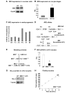
Figure 1
Figure 1. Mll2 oocyte-specific deletion mediated by the Gdf9-Cre allele leads to female infertility and premature follicle loss.
(A) RT-PCR analysis of Mll2 showed expression in ovarian granulosa cells (GC) and peri-ovulatory (GV) oocytes (Oo) as determined. (B) Real time PCR (QPCR) analysis of meiotically incompetent (immature) oocytes from 12-d-old females and mature peri-ovulatory (GV, germinal vesicle) oocytes showed a significant increase in Mll2 mRNA levels towards the peri-ovulatory stage (Student's t test, * p<0.05; three pools of oocytes were used in the analysis; n = 3). Results are shown relative to Mll2 levels in GV oocytes (RQ). Gapdh was used as endogenous control. (C) QPCR analysis of preimplantation embryos revealed that Mll2 is present in 1-cell embryos, likely as a maternal product, and also in subsequent embryonic stages up to the blastocyst (Bl) stage. Gapdh was used as endogenous control. Results are shown as arbitrary units (delta CT: CT values corrected by the endogenous control). Abbreviations: GV, germinal vesicle oocytes; 1-C, 1 cell embryos; 2-C, 2 cell embryos; 4-C, 4 cell embryos; M, morula; Bl, blatocyst. (D) cKO mice were generated by tissue-specific deletion of the Mll2 floxed (“F”) allele in the Mll2 null allele background. The “F” allele was generated after FLP recombination to remove the stop cassette. Cre recombination eliminates the second exon, thereby causing a frame-shift (“FC” allele). (E) Breeding scheme used to generate Mll2Gdf9 cKO mice. (F) Mll2Gdf9 cKO oocytes show a significant reduction in Mll2 mRNA levels (Student's t test, * p<0.05). Results are shown as means ± S.E. relative to WT (RQ) (three oocyte pools were used; n = 3), and Gapdh was used as endogenous control. (G) Mll2Gdf9 cKO oocytes show negligible levels of MLL2 protein as assessed by Western blot analysis; tubulin was used as loading control. (H) Mll2Gdf9 cKO females were infertile; the cumulative number of pups over a 6-mo period is shown (n = 10).
Figure 2
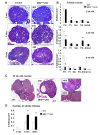
Figure 2. Mll2Gdf9 cKO females show increased ovarian follicular recruitment and loss.
(A) PAS-stained ovaries from 2-, 3-, and 8-wk-old mice; the number of follicles was greatly reduced in ovaries from 8-wk-old Mll2Gdf9 cKO mice. (B) Follicle counts from 2-, 3-, and 8-wk-old mice. At 2 wk, Mll2Gdf9 cKO ovaries showed significantly increased (ANOVA test, * p<0.05) primary follicles (Pr), whereas 3-wk-old Mll2Gdf9 cKO ovaries showed significantly reduced primordial follicles (Prl) and increased preantral follicles (PA) (ANOVA test, * p<0.05). Mll2Gdf9 cKO ovaries from 8-wk-old mice showed further reduction in Prl follicles (ANOVA test, * p<0.05). Means of follicle counts corrected by surface area ± S.E. are shown (ovarian sections from five individual females were used; n = 5). Abbreviations: Prl, primordial; Pr, primary; Sec, secondary; A, antral; CL, corpora lutea. (C) Mll2Gdf9 cKO ovaries from 36-wk-old mice stained with hematoxylin-eosin showed very few follicles (boxed area). (D) From 3 wk onwards, the number of atretic follicles was significantly higher in Mll2Gdf9 cKO ovaries, compared to controls. Means of follicle counts corrected by surface area ± S.E. are shown (ovarian sections from five individual females were used; n = 5). ANOVA test, * p<0.05.
Figure 3
Figure 3. Mll2Gdf9 cKO females show impaired ovulation.
(A) Ovulation rates were significantly reduced in 3-wk-old superovulated Mll2Gdf9 cKO females (Student's t test, ** p<0.01). Means ± S.E. are shown (five individual females were used; n = 5). (B) PAS-stained ovaries from superovulated 3-wk-old Mll2Gdf9 cKO mice showed trapped oocytes (yellow arrowheads), which correlated with decreased ovulation rates in this mouse line. (C) In vitro oocyte maturation studies; representative single plane confocal laser microscopy micrographs of peri-ovulatory oocytes cultured for 16 h to allow resumption of meiosis; arrows show lagging chromosomes. Magnification: 800×. Abbreviations: MI, meiosis I; MII, meiosis II; PB, polar body. (D) Percentages of meiosis I (MI), meiosis II (MII), or abnormal oocytes after 16 h in culture (ANOVA test, * p<0.05). Means ± S.E. of four independent experiments are shown; a total of 120 oocytes per genotype were analyzed.
Figure 4
Figure 4. Mll2Gdf9 cKO oocytes fail to establish transcriptional repression.
(A) Confocal laser microscopy analysis of peri-ovulatory oocytes stained with propidium iodide to visualize DNA (upper panel); oocytes were scored as displaying a surrounded nucleolus (SN) or a non-surrounded nucleolus (NSN) configuration (lower panel). Arrows show the nucleolus. No significant differences were observed between controls and Mll2Gdf9 cKO oocytes (ANOVA test, * p = 0.08). Means ± S.E. are shown; a total of 170 oocytes from five individual females were analyzed in three independent experiments. Magnification: 800×. (B) Run-on and confocal microscopy analyses of Br-UTP labeled nascent RNA in peri-ovulatory oocytes. Upper panel: Representative single plane confocal micrographs are shown as merge and split channels (Br-UTP, green staining; and DNA: propidium iodide, red staining). Lower panel: oocytes were scored as positive or negative for Br-UTP staining; note that Mll2Gdf9 cKO oocytes were transcriptionally active (ANOVA test, * p<0.05). Transcription was RNA Pol II dependent as it was abrogated by α-amanitin (αA) (magnification: 800×). Means ± S.E. are shown; a total of 90 oocytes from four individual females were evaluated in three independent experiments.
Figure 5
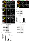
Figure 5. Mll2Gdf9 cKO oocytes show loss of bulk H3K4me2 and H3K4me3 as well as H4 hyperacetylation.
(A–C) Confocal microscopy micrographs showing H3K4 methylation levels in peri-ovulatory oocytes. (A) An increase in H3K4me1 is apparent in Mll2Gdf9 cKO oocytes (lower panel) compared to control (upper panel). In contrast, a decrease in H3K4me2 (B) and H3K4me3 (C) levels was observed in Mll2Gdf9 cKO oocytes (B and C, lower panels) compared to controls (B and C, upper panels). Magnification: 800×. Representative single plane micrographs are shown as merge and split channels of histone tail modifications (green) and DNA (red). (D–E) Western blot analysis of H3K4 methylation in chromatin fractions from peri-ovulatory oocytes. (D) Representative micrographs of Western blots showing H3K4me1, H3K4me2, and H3K4me3 levels. (E) Chemoluminescence quantification revealed a significant increase in global H3K4me1 and a significant decrease in global H3K4me2 and H3K4me3 levels in Mll2Gdf9 cKO oocytes; Student's t test, * p<0.05; ** p<0.01. Means ± S.E are shown. Three pools of 100 oocytes each from 3–4 females per genotype were used in three independent experiments; samples were normalized against total histones. (F) Confocal microscopy micrographs showing H4K12 acetylation levels in peri-ovulatory oocytes. An increase in H4K12 acetylation is apparent in Mll2Gdf9 cKO oocytes (lower panel) compared to controls (upper panel). Magnification: 800×. Representative single plane micrographs are shown as merge and split channels of histone tail modifications (green) and DNA (red). (G–H) Western blot analysis of H4K12 acetylation (H3K12(ac)) in chromatin fractions from peri-ovulatory oocytes. (G) Representative micrographs of Western blots showing H4K12 acetylation levels. (H) Chemoluminescence quantification revealed a significant increase in global H4K12 acetylation in Mll2Gdf9 cKO oocytes; Student's t test, * p<0.05; Means ± S.E are shown. Three pools of 100 oocytes each from 3–4 females per genotype were used in three independent experiments; samples were normalized against total histones. (I) Western blot analysis of other histone tail modifications in chromatin fractions from peri-ovulatory oocytes. Representative micrographs of Western blots showing H3K9me2, H3K27me3, H4K20me1, and H3(ac); total histones were used as internal loading controls. No changes were observed in the levels of any of the histone tail modifications analyzed. Three pools of 100 oocytes each from 3–4 females per genotype were used in three independent experiments.
Figure 6
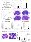
Figure 6. Oocyte-specific deletion of Mll2 mediated by the Zp3-Cre allele.
(A) Breeding scheme used to generate Mll2Zp3cKO mice. (B) Zp3-cKO oocytes show a significant reduction in Mll2 mRNA levels (Student's t test, * p<0.05). Results are shown as means ± S.E. relative to WT (RQ) (three oocyte pools were used; n = 3) and Gapdh was used as endogenous control. (C) FSH levels were significantly higher in serum samples from 8-wk-old Mll2Zp3 cKO females compared to controls (Student's t test, * p<0.05; n = 10). (D) Fertility studies shown as cumulative number of pups over a 6-mo period. Mll2Zp3cKO females were infertile. (G) Ovulation rates were significantly reduced in Mll2Zp3cKO females compared to controls (WT) (Student's t test; * p<0.05; n = 5). (E) PAS-stained ovaries from 3- and 36-wk-old mice. Original magnification: 25× and 50×; note that different from Mll2Gdf9 cKO mice, Mll2Zp3 cKO ovaries still contain follicles at 36 wk of age. (F) Follicle counts in Mll2Zp3 cKO ovaries. Mll2Zp3 cKO ovaries had a significantly lower number of primordial follicles and increased numbers of secondary and multilayer preantral follicles 3 wk of age (left panel). By 8 wk of age (right panel), the number of primordial and multilayer preantral follicles is significantly reduced and the number of atretic follicles is increased in Mll2Zp3 cKO ovaries. (ANOVA test; * p<0.05; ovarian sections from five females were used in the analysis; n = 5). Abbreviations: Prl, primordial; Pr, primary; Sec, secondary; PA, preantral; A, antral; CL, corpora lutea; Atr, atretic. (I) Cultures of embryos from Mll2Zp3 cKO females crossed to WT males showed a developmental arrest between the 1-cell and the 4-cell stages. Results are presented as the % embryos (average) ± standard error from five independent experiments. ANOVA test; * p<0.05. A total of 95 embryos from six females per genotype were used in three independent experiments. Abbreviations: C, cell; Frag, fragmented; WT, control wild type.
Figure 7
Figure 7. Mll2Zp3 cKO oocytes show decreased H3K4 tri-methylation.
(A–B) Confocal microscopy micrographs showing H3K4 methylation levels in peri-ovulatory oocytes. (A) An increase in H3K4me1 is apparent in Mll2Zp3 cKO oocytes (lower panel) compared to control (upper panel). In contrast, a decrease in H3K4me3 (B) levels was observed in Mll2Zp3 cKO oocytes (lower panel) compared to controls (upper panel). Magnification: 800×. Representative single plane micrographs are shown as merge and split channels of histone tail modifications (green) and DNA (red). (C) Representative micrographs of Western blots showing H3K4me3 levels in chromatin fractions from Mll2Zp3 cKO peri-ovulatory oocytes. A decrease in global H3K4me3 is apparent in Mll2Zp3c KO oocytes (Student's t test, * p<0.05). Three pools of 100 oocytes each from 3–4 females per genotype were used in three independent experiments. Total histones were used as internal loading controls.
Figure 8
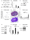
Figure 8. MLL2 is not required for granulosa cell function.
(A) Breeding scheme used to generate granulosa cell-specific cKO mice (Mll2Amhr2 cKO). (B) QPCR analysis of Mll2 levels in granulosa cells from wild type (WT), Mll2F/ (heterozygous control), and Mll2Amhr2 cKO females (granulosa cells from three individual females were used in the analysis; n = 3). Mll2Amhr2 cKO granulosa cells show a significant decrease in the levels of Mll2, whereas Mll2F/ granulosa cells show the expected 50% decrease in Mll2 levels (ANOVA test; * p<0.05). Gadpdh was used as internal control. (C) Western blot analysis of Mll2Amhr2 cKO granulosa cells showed decreased MLL2 protein levels. Tubulin was used as loading control. (D) Fertility studies shown as cumulative number of pups over a 6-mo period. Mll2Amhr2 cKO females displayed a small reduction in the number of pups (n = 10). (E) PAS-stained ovaries from 36-wk-old Mll2Amhr2 cKO mice showed PAS-positive zona pellucida remnants (oocyte remnants, arrowheads). Note that the number of follicles present at this age is higher than that observed in the oocyte-specific cKO mouse lines. (F) Follicle counts in 36-wk-old ovaries. Mll2Amhr2 cKO ovaries show a significant decrease in the number of quiescent amd growing follicles and a significantly higher number of atretic follicles (ovarian sections from five females were used in this study; n = 5). Abbreviations: Prl, primordial; Pr, primary; Sec, secondary; PA, preantral; A, antral; CL, corpora lutea. ANOVA test; * p<0.05. (G–H) Western blot analysis of H3K4 methylation in chromatin fractions from granulosa cells. (G) Representative micrographs of Western blots showing H3K4me1, H3K4me2, and H3K4me3 levels. (H) Chemoluminescence quantification revealed a significant decrease in global H3K4me3 levels in Mll2Amhr2 cKO granulosa cells (Student's t test, * p<0.05). Means ± S.E are shown. Granulosa cells from three individual females per genotype were used in three independent experiments; samples were normalized against total histones. (G) Representative Western blot analysis of the repressive mark H3K27me3 showed no global changes in Mll2Amhr2 cKO granulosa cells. Total histones were used as internal loading control.
Figure 9
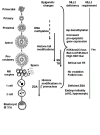
Figure 9. Summary of MLL2 function during gametogenesis and early embryogenesis.
During normal folliculogenesis, oocytes are transcriptionally active and grow in size but do not proliferate. In contrast, the surrounding somatic cells are actively dividing. A number of epigenetic marks are acquired by oocytes during folliculogenesis, including DNA methylation and histone tail modifications. Towards the peri-ovulatory stage, oocytes undergo a large chromatin re-arrangenment into the surrounded nucleolus (SN) configuration and global transcriptional repression (TR). Upon ovulation and fertilization, the maternal pronucleus retains the majority of the histone tail modification marks established in the oocyte, whereas the male pronucleus acquires them progressively. Between the late 1-cell and the 2-cell embryo stages, the maternal mRNA and protein stores are depleted and the zygotic genome is activated (ZGA). MLL2 is expressed in oocytes and during pre-implantation development. Mll2 oocyte-specific cKO females are infertile due to anovulation and follicle loss. Mll2-deficient oocytes show reduced H3K4me3 but not H3K4me1, indicating another enzyme monomethylates H3K4 in oocytes. Oocytes lacking Mll2 also show abnormal expression of pro-apoptotic genes and Iap elements, which may contribute to oocyte death and, ultimately, follicle loss. Although Mll2-deficient oocytes display the SN configuration, they fail to repress transcription, likely due to loss of communication with the granulosa cell compartment. Finally, studies in the hypomorph and the Mll2Zp3 cKO model reveal that MLL2 is required either directly or indirectly for embryogenesis and ZGA. Together the results suggest that MLL2 is continuously required during oogenesis and early embryonic development.

Similar articles

Cited by

References

    1. De La Fuente R, Viveiros M. M, Burns K. H, Adashi E. Y, Matzuk M. M, et al. Major chromatin remodeling in the germinal vesicle (GV) of mammalian oocytes is dispensable for global transcriptional silencing but required for centromeric heterochromatin function. Dev Biol. 2004;275:447–458. - PubMed
    1. Reik W, Santos F, Mitsuya K, Morgan H, Dean W. Epigenetic asymmetry in the mammalian zygote and early embryo: relationship to lineage commitment? Philos Trans R Soc Lond B Biol Sci. 2003;358:1403–1409; discussion 1409. - PMC - PubMed
    1. Farthing C. R, Ficz G, Ng R. K, Chan C. F, Andrews S, et al. Global mapping of DNA methylation in mouse promoters reveals epigenetic reprogramming of pluripotency genes. PLoS Genet. 2008;4:e1000116. doi: 10.1371/journal.pbio.1000116. - DOI - PMC - PubMed
    1. Silva J, Nichols J, Theunissen T. W, Guo G, van Oosten A. L, et al. Nanog is the gateway to the pluripotent ground state. Cell. 2009;138:722–737. - PMC - PubMed
    1. Nichols J, Smith A. Naive and primed pluripotent states. Cell Stem Cell. 2009;4:487–492. - PubMed

Publication types

  NODES
INTERN 4
Note 3
twitter 2