Integrative modeling of the cardiac ventricular myocyte
- PMID: 20865780
- PMCID: PMC3110595
- DOI: 10.1002/wsbm.122
Integrative modeling of the cardiac ventricular myocyte
Abstract
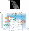
Cardiac electrophysiology is a discipline with a rich 50-year history of experimental research coupled with integrative modeling which has enabled us to achieve a quantitative understanding of the relationships between molecular function and the integrated behavior of the cardiac myocyte in health and disease. In this paper, we review the development of integrative computational models of the cardiac myocyte. We begin with a historical overview of key cardiac cell models that helped shape the field. We then narrow our focus to models of the cardiac ventricular myocyte and describe these models in the context of their subcellular functional systems including dynamic models of voltage-gated ion channels, mitochondrial energy production, ATP-dependent and electrogenic membrane transporters, intracellular Ca dynamics, mechanical contraction, and regulatory signal transduction pathways. We describe key advances and limitations of the models as well as point to new directions for future modeling research. WIREs Syst Biol Med 2011 3 392-413 DOI: 10.1002/wsbm.122
Copyright © 2010 John Wiley & Sons, Inc.
Figures


Similar articles
-
Stretch-activated current in human atrial myocytes and Na+ current and mechano-gated channels' current in myofibroblasts alter myocyte mechanical behavior: a computational study.Biomed Eng Online. 2019 Oct 25;18(1):104. doi: 10.1186/s12938-019-0723-5. Biomed Eng Online. 2019. PMID: 31653259 Free PMC article.
-
Computational Modeling of Cardiac Electrophysiology.Methods Mol Biol. 2024;2735:63-103. doi: 10.1007/978-1-0716-3527-8_5. Methods Mol Biol. 2024. PMID: 38038844
-
A computational model integrating electrophysiology, contraction, and mitochondrial bioenergetics in the ventricular myocyte.Biophys J. 2006 Aug 15;91(4):1564-89. doi: 10.1529/biophysj.105.076174. Epub 2006 May 5. Biophys J. 2006. PMID: 16679365 Free PMC article.
-
Cardiac systems biology and parameter sensitivity analysis: intracellular Ca2+ regulatory mechanisms in mouse ventricular myocytes.Adv Biochem Eng Biotechnol. 2008;110:25-45. doi: 10.1007/10_2007_093. Adv Biochem Eng Biotechnol. 2008. PMID: 18437298 Review.
-
Dynamic clamp: a powerful tool in cardiac electrophysiology.J Physiol. 2006 Oct 15;576(Pt 2):349-59. doi: 10.1113/jphysiol.2006.115840. Epub 2006 Jul 27. J Physiol. 2006. PMID: 16873403 Free PMC article. Review.
Cited by
-
Calcium Sparks in the Heart: Dynamics and Regulation.Res Rep Biol. 2015;6:203-214. doi: 10.2147/RRB.S61495. Epub 2015 Oct 16. Res Rep Biol. 2015. PMID: 27212876 Free PMC article.
-
Cell-specific models of hiPSC-CMs developed by the gradient-based parameter optimization method fitting two different action potential waveforms.Sci Rep. 2024 Jun 7;14(1):13086. doi: 10.1038/s41598-024-63413-0. Sci Rep. 2024. PMID: 38849433 Free PMC article.
-
Calibration of ionic and cellular cardiac electrophysiology models.Wiley Interdiscip Rev Syst Biol Med. 2020 Jul;12(4):e1482. doi: 10.1002/wsbm.1482. Epub 2020 Feb 21. Wiley Interdiscip Rev Syst Biol Med. 2020. PMID: 32084308 Free PMC article. Review.
-
Exploiting mathematical models to illuminate electrophysiological variability between individuals.J Physiol. 2012 Jun 1;590(11):2555-67. doi: 10.1113/jphysiol.2011.223313. Epub 2012 Apr 10. J Physiol. 2012. PMID: 22495591 Free PMC article. Review.
-
Estimating the probability of early afterdepolarizations and predicting arrhythmic risk associated with long QT syndrome type 1 mutations.Biophys J. 2023 Oct 17;122(20):4042-4056. doi: 10.1016/j.bpj.2023.09.001. Epub 2023 Sep 12. Biophys J. 2023. PMID: 37705243 Free PMC article.
References
-
- Cai D, Winslow RL, Noble D. Effects of gap junction conductance on dynamics of sinoatrial node cells: two-cell and large-scale network models. IEEE Trans Biomed Eng. 1994;41:217–231. - PubMed
-
- Soeller C, Cannell MB. Examination of the transverse tubular system in living cardiac rat myocytes by 2-photon microscopy and digital image-processing techniques. Circ Res. 1999;84:266–275. - PubMed
-
- Bers DM. Cardiac excitation–contraction coupling. Nature. 2002;415:198–205. - PubMed
-
- Forbes MS, Sperelakis N. Association between mitochondria and gap junctions in mammalian myocardial cells. Tissue Cell. 1982;14:25–37. - PubMed
-
- Shiels HA, White E. The Frank-Starling mechanism in vertebrate cardiac myocytes. J Exp Biol. 2008;211:2005–2013. - PubMed
Publication types
MeSH terms
Grants and funding
LinkOut - more resources
Full Text Sources
Other Literature Sources

