. 2010 Oct 6;30(40):13305-13.
doi: 10.1523/JNEUROSCI.3010-10.2010.

Histone H1 poly[ADP]-ribosylation regulates the chromatin alterations required for learning consolidation

Affiliations

Histone H1 poly[ADP]-ribosylation regulates the chromatin alterations required for learning consolidation

Angela Fontán-Lozano et al. J Neurosci. .

Abstract

Memory formation requires changes in gene expression, which are regulated by the activation of transcription factors and by changes in epigenetic factors. Poly[ADP]-ribosylation of nuclear proteins has been postulated as a chromatin modification involved in memory consolidation, although the mechanisms involved are not well characterized. Here we demonstrate that poly[ADP]-ribose polymerase 1 (PARP-1) activity and the poly[ADP]-ribosylation of proteins over a specific time course is required for the changes in synaptic plasticity related to memory stabilization in mice. At the molecular level, histone H1 poly[ADP]-ribosylation was evident in the hippocampus after the acquisition period, and it was selectively released in a PARP-1-dependent manner at the promoters of cAMP response element-binding protein and nuclear factor-κB dependent genes associated with learning and memory. These findings suggest that histone H1 poly[ADP]-ribosylation, and its loss at specific loci, is an epigenetic mechanism involved in the reprogramming of neuronal gene expression required for memory consolidation.


Figures
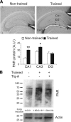
Figure 1.
Figure 1.
Synthesis of the PAR polymer is induced after object recognition training. A, The levels of PAR polymer were determined by immunohistochemistry in brains from mice killed 15 min after a 15 min OR training session (n = 5 mice per group). Bar chart illustrating the quantification of PAR polymer immunoreactivity in the CA1, CA3, and dentate gyrus (DG) of the hippocampus in habituated (white bars) and trained (black bars) mice. B, Representative Western blot probed for protein poly[ADP]-ribosylation in the hippocampus of mice administered the PARP-1 inhibitor Tiq-A or the vehicle alone and killed 15 min after a 15 min OR training session. Densitometry of three independent experiments is shown. Asterisks indicate significant differences in the densitometry between habituated and trained mice; *p < 0.05; **p < 0.01.
Figure 2.
Figure 2.
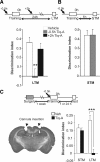
Hippocampal PARP-1 activation is required for object recognition memory consolidation. A, B, Mice were subjected to a 15 min OR training session, and 1 or 24 h after training, their STM and LTM, respectively, was tested. To test the role of the PAR polymer on memory consolidation, either 0.5 mg/kg Tiq-A or saline was injected at varying times from the end of the training session (from −0.5 to +2 h). The bar chart illustrates the discrimination indices calculated as (tnoveltfamiliar)/(tnovel + tfamiliar) during LTM (A) and STM (B) sessions (n = 8 for each group). The bars in white represent saline administration, and gray and black bars represent the different times of Tiq-A administration. C, The effects of Tiq-A infusion directly into the hippocampus of mice. Tiq-A was administered 5 min before a 15 min training session, and the STM and LTM were assayed. Left, Representative microphotograph of the cannula located in the hippocampus. Right, The bar chart illustrates the discrimination indices during STM and LTM (n = 10 mice per group). **p < 0.01; ***p < 0.001.
Figure 3.
Figure 3.
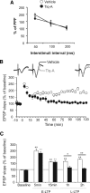
PARP-1 inhibition blocked long-lasting changes in synaptic efficacy. A, Basal excitatory neurotransmission was measured using paired-pulse facilitation with interpulse intervals from 50 to 200 ms in the presence or absence of Tiq-A. The percentage of paired-pulse facilitation as a function of the interpulse interval in mice receiving Tiq-A or the vehicle alone (white circle, vehicle; black circle, Tiq-A mice; n = 6 for each group) is presented. B, Changes in the slope of the fEPSP induced by six HFS [5 trains (200 Hz, 100 ms) of pulses at a rate of 1/s]. Note that the evoked long-lasting LTP lasted for up to 2 h in the vehicle-administered mice (white) but not in Tiq-A-administered mice (black; n = 6 for each group). Representative recordings at baseline and 15 or 120 min after HF from mice that received Tiq-A or the vehicle alone. C, Summary of the changes in fEPSP slope at different times after six HFS trains: baseline, 5 and 15 min after HFS [early LTP (E-LTP)], and 1 and 2 h after HFS [long-lasting LTP (L-LTP)]. All data are shown as mean values ± SEM. *p < 0.05 and **p < 0.01 indicates statistically significant difference between the vehicle- and Tiq-A-injected mice at different times after HFS.
Figure 4.
Figure 4.
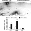
The HFS protocol used for LTP induces an increase in PAR polymer in the hippocampus. The levels of PAR polymer were determined by immunohistochemistry in brains from mice killed 1 h after the application of six HFS stimulations to the entorhinal cortex (n = 3 mice per group). Bar chart illustrating the quantification of PAR polymer immunoreactivity in the CA1, CA3, and dentate gyrus (DG) of unstimulated (white bars) and stimulated (black bars) hippocampi. Significant differences of the data from unstimulated and stimulated slices for each hippocampal region. **p < 0.01.
Figure 5.
Figure 5.
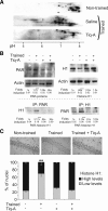
OR training sessions induced histone H1 poly[ADP]-ribosylation and protein decrease in the hippocampus. A, Protein extracts from the hippocampus obtained from untrained mice and those that had just finished their training, in the presence or absence of Tiq-A (0.5 mg/kg), were analyzed by two-dimensional electrophoresis, and histone H1 immunoreactivity was determined. Poly[ADP]-ribosylation of histone H1 is reflected by a shift in pI toward acidic pH values. B, Hippocampal protein extracts from untrained and trained mice that received Tiq-A (0.5 mg/kg) or the vehicle alone and were killed 15 min after training session were used to immunoprecipitate PAR polymer or histone H1. The immunoprecipitates were examined in Western blots probed for histone H1 and PAR polymer (bottom). As an internal control, 10% of the protein used for the immunoprecipitation assays was examined in Western blots probed for PAR polymer, histone H1, and actin (top). Values illustrate the densitometric quantification of PAR polymer and histone H1 immunoreactivity in each experimental group or the relative amount of histone H1 poly[ADP]-ribosylated (PAR-histone H1) in each experimental group with respect to the untrained animals. C, Immunohistochemistry of histone H1 expression in the hippocampal CA1 field of mice killed 1 h after training or manipulation in the presence or absence of Tiq-A. The top shows representative images of histone H1 in CA1 neurons after the manipulations. The bottom shows a bar graph of the percentage of neurons labeled with high or low amounts of histone H1 under the different conditions (n = 5 mice per groups). IP, Immunoprecipitation. **p < 0.01; ***p < 0.001.
Figure 6.
Figure 6.
OR training promotes PARP-1-dependent histone H1 clearance and RNA pol II recruitment to NF-κB and CREB gene promoters. A, Semiquantitative reverse transcription-PCR analysis of i-nos, tnf-α, c-fos, c-jun, and egr-1 mRNA from the hippocampus of untrained mice and from that obtained 1 h after OR training in mice that received Tiq-A or the vehicle alone. GAPDH mRNA served as an internal control. The bar graph represents the trained/untrained ratio of the normalized values of gene expression in the presence or absence of Tiq-A (white and black bars, respectively; n = 5 mice per group). B, C, The levels of histone H1 (B) and RNA pol II (C) at the tnf-α, c-fos, c-jun, and egr-1 promoters were studied by chromatin immunoprecipitation in hippocampal chromatin complexes obtained from untrained and trained mice that received Tiq-A or the vehicle alone. Histograms represent the trained/untrained ratio of normalized values of gene promoter amplification in the presence or absence of Tiq-A (black and white bars, respectively; n = 3 mice per group). Asterisks above the line indicate a difference between the two treatments (vehicle and Tiq-A), whereas asterisks above a bar graph indicate a significant difference between trained and untrained mice. *p < 0.05; **p < 0.01; ***p < 0.001.

Similar articles

Cited by

References

    1. Abel T, Kandel E. Positive and negative regulatory mechanisms that mediate long-term memory storage. Brain Res Rev. 1998;26:360–378. - PubMed
    1. Abel T, Lattal KM. Molecular mechanisms of memory acquisition, consolidation and retrieval. Curr Opin Neurobiol. 2001;11:180–187. - PubMed
    1. Aguilar-Quesada R, Muñoz-Gámez JA, Martín-Oliva D, Peralta-Leal A, Quiles-Pérez R, Rodríguez-Vargas JM, Ruiz de Almodóvar M, Conde C, Ruiz-Extremera A, Oliver FJ. Modulation of transcription by PARP-1: consequences in carcinogenesis and inflammation. Curr Med Chem. 2007;14:1179–1187. - PubMed
    1. Ahn HJ, Hernandez CM, Levenson JM, Lubin FD, Liou HC, Sweatt JD. c-Rel, an NF-kappaB family transcription factor, is required for hippocampal long-term synaptic plasticity and memory formation. Learn Mem. 2008;15:539–549. - PMC - PubMed
    1. Alarcón JM, Malleret G, Touzani K, Vronskaya S, Ishii S, Kandel ER, Barco A. Chromatin acetylation, memory, and LTP are impaired in CBP+/− mice: a model for the cognitive deficit in Rubinstein-Taybi syndrome and its amelioration. Neuron. 2004;42:947–959. - PubMed

Publication types

MeSH terms

LinkOut - more resources

  NODES
admin 6
INTERN 2
Note 1
twitter 2