. 2010 Oct 19;18(4):382-95.
doi: 10.1016/j.ccr.2010.08.010.

Pharmacologic inhibition of the anaphase-promoting complex induces a spindle checkpoint-dependent mitotic arrest in the absence of spindle damage

Affiliations

Pharmacologic inhibition of the anaphase-promoting complex induces a spindle checkpoint-dependent mitotic arrest in the absence of spindle damage

Xing Zeng et al. Cancer Cell. .

Abstract

Microtubule inhibitors are important cancer drugs that induce mitotic arrest by activating the spindle assembly checkpoint (SAC), which, in turn, inhibits the ubiquitin ligase activity of the anaphase-promoting complex (APC). Here, we report a small molecule, tosyl-L-arginine methyl ester (TAME), which binds to the APC and prevents its activation by Cdc20 and Cdh1. A prodrug of TAME arrests cells in metaphase without perturbing the spindle, but nonetheless the arrest is dependent on the SAC. Metaphase arrest induced by a proteasome inhibitor is also SAC dependent, suggesting that APC-dependent proteolysis is required to inactivate the SAC. We propose that mutual antagonism between the APC and the SAC yields a positive feedback loop that amplifies the ability of TAME to induce mitotic arrest.


Conflict of interest statement

The authors declare no financial conflict of interest.

Figures
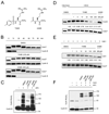
Figure 1
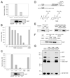
Figure 1. TAME inhibits APC activation by perturbing binding of Cdc20 or Cdh1
(A) Structures of TAME and AAME. (B) TAME induces mitotic arrest in Xenopus extract. Recombinant cyclin B1/Cdk1 was added to interphase extract in the presence of compounds. Cdc27 phosphorylation and cyclin B1 levels were examined by immunoblot. (C) TAME inhibits APC activation. Compounds were added to mitotic Xenopus extract immediately before APC immunoprecipitation. The activity of the isolated APC was measured in a reconstituted assay. (D) TAME inhibits Cdc20 association with mitotic APC. Compounds were added to mitotic Xenopus extract prior to APC immunoprecipitation. Numbers represent CCD-imaging based-intensity quantitation of the immunoblot, and show the relative amount of Cdc20 normalized to Cdc27. (E) TAME inhibits Cdh1 association with interphase APC. Interphase Xenopus extract was pre-incubated with compounds for 30 min prior to adding recombinant Cdh1 and APC immunoprecipitation. (F) TAME inhibits APC activation by Cdh1. Interphase Xenopus extract was pre-incubated with compound for 30 min prior to adding recombinant Cdh1 and APC immunoprecipitation. The activity of the isolated APC was measured in a reconstituted assay. See also Figure S1.
Figure 2
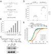
Figure 2. TAME binds to the APC and inhibits binding of the IR tail of activator proteins
(A) TAME binds Xenopus APC. 3H-TAME was added to interphase extract or to extract that had been partially or completely immunodepleted of APC. Remaining APC was then immunoprecipitated and the associated radioactivity was measured by scintillation counting. Residual APC levels were measured by immunoblot with Cdc27 antibody. Specific binding was calculated as described in the methods. (B) Unlabeled TAME competes with 3H-TAME for binding to Xenopus APC. 3H-TAME was added to interphase extract with unlabeled TAME or AAME prior to APC immunoprecipitation. (C) 3H-TAME binds to human APC. The experiment in 2A was repeated with lysate from asynchronous HeLa cells. (D) Schematic of Cdc20, the C-box containing fragment, and structures of the IR tail and TAME. (E) TAME inhibits the interaction between the Cdh1 C-terminal IR peptide and the APC. Left: Resin coupled with cysteine (Ctrl resin), Cdh1 C-terminal peptide (WT), or the peptide lacking the C-terminal isoleucine and arginine (ΔIR) was incubated with interphase Xenopus extract, washed, and the amount of bound Cdc27 was analyzed by immunoblot. Right: Cdh1 C-terminal resin was incubated with interphase extract in the presence of compounds and the amount of Cdc27 was analyzed as above. (F) TAME does not inhibit the interaction between the C-box and the APC. A 159-amino acid N-terminal fragment of Cdc20 containing the C-box fused to GST (GST-CDC20 N159 WT) or the same fragment lacking the C-box (GST-CDC20 N159 ΔC-box) were bound to glutathione resin and incubated with mitotic Xenopus extract in the presence of compounds. Bound Cdc27 was analyzed by immunoblot. (G) TAME inhibits IR-peptide crosslinking to APC subunits. Purified interphase Xenopus APC was incubated with an IR peptide coupled to a biotin-containing label-transfer reagent, in the presence or absence of compounds, prior to photocrosslinking. Reaction products were detected by streptavidin-HRP. See also Figure S2.
Figure 3
Figure 3. TAME inhibits binding of wild type Cdc20 to the APC, but not binding of a ΔIR mutant
Mitotic APC immunoprecipitated from Xenopus extract was washed with XB high salt (500 mM KCl) and XB to remove endogenous Cdc20 prior to incubation with in vitro translated wild type Xenopus Cdc20 or the ΔIR mutant. Various competitors were added during incubation as indicated. Unbound proteins were washed away and bound Cdc20 was analyzed by immunoblot. Numbers represent the amount of Cdc20 normalized to Cdc27.
Figure 4
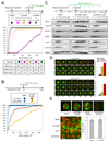
Figure 4. ProTAME inhibits APC activity in Xenopus extract and inhibits Cdh1-dependent APC activity during interphase in HeLa cells
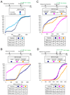
(A) Structures of proTAME and proAAME. (B) ProTAME inhibits cyclin B-luciferase degradation in mitotic Xenopus extract. Different concentrations of proTAME or proAAME were added to mitotic Xenopus extract containing cyclin B-luciferase reporter. Samples were collected at 60 min and the remaining reporter level was measured by luminescence. (C) ProTAME blocks Cdh1 association with the APC. HeLa cells were released from nocodazole and treated with proTAME in G1. APC was immunoprecipitated from cell lysates and the amount of Cdc27 and Cdh1 was analyzed by immunoblot. (D) ProTAME restores mitotic entry in Emi1-depleted cells. HeLa cells were transfected with control siRNA or Emi1 siRNA and treated with DMSO or proTAME 24 h after transfection and then imaged for 48 h. About 400 cells were analyzed in each experiment, and the proportion that failed to enter mitosis during the 48 h of imaging was calculated. Results of 3 independent experiments are shown. Statistical significance was calculated using an unpaired t-test. (E) ProTAME causes a mitotic entry delay if added during S-phase. HeLa H2B-GFP cells were released from a double thymidine block and proTAME (12 µM) was added at different time points as indicated. Mitotic entry was monitored by time-lapse imaging. Cumulative frequency curves of the time of mitotic entry are shown. Statistical analysis, including mean, median, statistical significance and number of cells analyzed per condition for all experiments is included in Table S1. See also Figure S3.
Figure 5
Figure 5. ProTAME induces mitotic arrest without disrupting the mitotic spindle
(A) ProTAME induces mitotic arrest in HeLa cells. Double thymidine synchronized HeLa H2B-GFP cells were treated with compounds and analyzed by time-lapse imaging. Cumulative frequency curves of mitotic duration and cell fate distributions are shown. (B) Partial Cdc20 knockdown sensitizes cells to proTAME treatment. Asynchronous HeLa H2B-GFP cells were transfected with control or Cdc20 siRNA 24 h prior to treatment with compounds. (C) ProTAME stabilizes endogenous APC substrates. Double thymidine synchronized HeLa cells were treated with compounds. (D) ProTAME stabilizes exogenous cyclin B1-GFP and cyclin A2-GFP in HeLa cells. HeLa H2B-RFP cells transduced with cyclin-GFP adenoviruses were treated with 20 µM proTAME or proAAME, or 150 nM nocodazole. Bar: 12 µm. Representative cells are shown. For quantitation, the fraction of GFP intensity remaining at 60 min as compared to the onset of mitosis was determined (n≥ 30 individual cells per treatment). Error bars represent standard error of the mean. (E) ProTAME does not disrupt mitotic spindles or alter interkinetochore distance. Asynchronous HeLa cells were treated with compounds for 2 h, and then stained with anti-tubulin (green) and CREST (red) antibody. Representative images are shown. Bar: 3 µm. Representative images of kinetochore pairs are shown. Bar: 1.2 µm. Inter-kinetochore distance was measured in DMSO or proTAME treated cells (n=55, p=0.23). Error bars represent standard deviation. See also Figure S4 and Movies S1–4.
Figure 6
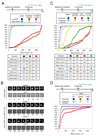
Figure 6. ProTAME-induced mitotic arrest is SAC-dependent
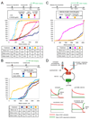
(A) ProTAME-induced mitotic arrest is Mad2-dependent. HeLa H2B-GFP cells were transfected with indicated siRNAs between rounds of thymidine treatment. Following release, cells were treated with compounds and analyzed by time-lapse imaging. A graph of the same data with an expanded x-axis is shown in Figure S5A. (B) ProTAME rescues the mitotic defect induced by Mad2 knockdown. Asynchronous HeLa H2B-GFP cells were treated with Mad2 siRNA 24 h prior to addition of compound. Bar: 10 µm. (C) ProTAME-induced mitotic arrest is hesperadin-sensitive. Double thymidine synchronized HeLa H2B-GFP cells were treated with compounds 8 h following release. (D) UbcH10 or Cdc27 knockdown induces a hesperadin-sensitive mitotic delay. HeLa H2B-GFP cells were transfected with indicated siRNA between rounds of thymidine synchronization and treated with hesperadin 8 h following release. See also Figure S5 and Movies S5–6.
Figure 7
Figure 7. MG132-induced mitotic arrest is SAC-dependent
(A) MG132-induced arrest is Mad2-dependent. HeLa H2B-GFP cells were transfected with indicated siRNAs between rounds of thymidine synchronization, treated with compounds, and followed by time-lapse imaging. (B) MG132-induced arrest is hesperadin-sensitive, but mitotic exit can be suppressed by proTAME. Double thymidine synchronized HeLa cells were treated with compounds. (C) Taxol cannot restore mitotic arrest in the presence of MG132 and hesperadin. Double thymidine synchronized HeLa cells were treated with compounds. (D) Mitotic arrest induced by a higher concentration of MG132 remains hesperadin-sensitive. Double thymidine synchronized HeLa cells were treated with compounds. M: MG132; pT: proTAME; H: hesperadin. See also Figure S6.
Figure 8
Figure 8. Microtubule inhibitors require protein synthesis for mitotic arrest whereas proTAME and MG132 do not
(A) Mitotic arrest induced by microtubule inhibitors requires protein synthesis but proTAME-induced arrest does not. Double thymidine synchronized HeLa-H2B-GFP cells were treated with compounds and followed by time-lapse imaging. CHX: cycloheximide. (B) MG132 (10 µM)-induced arrest is cycloheximide-resistant but Mad2-dependent. HeLa cells were transfected with indicated siRNAs between rounds of thymidine synchronization. (C) MG132 (10 µM)-induced arrest is cycloheximide-resistant but hesperadin-sensitive. Double thymidine synchronized HeLa cells were treated with compounds. (D) Model. In the bottom panels, the x-axis indicates time from mitotic entry. See also Figure S7.

Similar articles

Cited by

References

    1. Bekier ME, Fischbach R, Lee J, Taylor WR. Length of mitotic arrest induced by microtubule-stabilizing drugs determines cell death after mitotic exit. Mol Cancer Ther. 2009;8:1646–1654. - PubMed
    1. Biggins S, Murray AW. The budding yeast protein kinase Ipl1/Aurora allows the absence of tension to activate the spindle checkpoint. Genes Dev. 2001;15:3118–3129. - PMC - PubMed
    1. Brito DA, Rieder CL. Mitotic checkpoint slippage in humans occurs via cyclin B destruction in the presence of an active checkpoint. Curr Biol. 2006;16:1194–1200. - PMC - PubMed
    1. Brito DA, Yang Z, Rieder CL. Microtubules do not promote mitotic slippage when the spindle assembly checkpoint cannot be satisfied. J Cell Biol. 2008;182:623–629. - PMC - PubMed
    1. Burton JL, Tsakraklides V, Solomon MJ. Assembly of an APC-Cdh1-substrate complex is stimulated by engagement of a destruction box. Mol Cell. 2005;18:533–542. - PubMed

Publication types

MeSH terms

  NODES
Association 3
twitter 2