. 2011 Jan;85(2):1086-98.
doi: 10.1128/JVI.00877-10. Epub 2010 Nov 3.

Modification of nonstructural protein 1 of influenza A virus by SUMO1

Affiliations

Modification of nonstructural protein 1 of influenza A virus by SUMO1

Ke Xu et al. J Virol. 2011 Jan.

Abstract

Nonstructural protein 1 (NS1) is one of the major factors resulting in the efficient infection rate and high level of virulence of influenza A virus. Although consisting of only approximately 230 amino acids, NS1 has the ability to interfere with several systems of the host viral defense. In the present study, we demonstrate that NS1 of the highly pathogenic avian influenza A/Duck/Hubei/L-1/2004 (H5N1) virus interacts with human Ubc9, which is the E2 conjugating enzyme for sumoylation, and we show that SUMO1 is conjugated to H5N1 NS1 in both transfected and infected cells. Furthermore, two lysine residues in the C terminus of NS1 were identified as SUMO1 acceptor sites. When the SUMO1 acceptor sites were removed by mutation, NS1 underwent rapid degradation. Studies of different influenza A virus strains of human and avian origin showed that the majority of viruses possess an NS1 protein that is modified by SUMO1, except for the recently emerged swine-origin influenza A virus (S-OIV) (H1N1). Interestingly, growth of a sumoylation-deficient WSN virus mutant was retarded compared to that of wild-type virus. Together, these results indicate that sumoylation enhances NS1 stability and thus promotes rapid growth of influenza A virus.


Figures
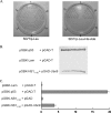
FIG. 1.
FIG. 1.
A/Duck/Hubei/L-1/2004 (H5N1) NS1 is a new human Ubc9-interacting protein. (A) Yeast AH109 cells cotransformed with the indicated combinations of bait and prey plasmids were grown on SD/Trp-Leu medium to select for double-transformed cells (left) and then further grown on an SD/Trp-Leu-His-Ade plate for yeast two-hybrid interaction screening (right). 1, negative control (pGBK-Lam+pGAD-T); 2, positive control (pGBK-p53+pGAD-T); 3, pGBK+pGAD; 4, pGBK+pGAD-Ubc9; 5, pGBK-NS11-162+pGAD; 6, pGBK-NS11-162+ pGAD-Ubc9. (B) Colony lift filter assay. The indicated plasmid pairs were cotransfected into the yeast strain SFY526. Colonies grown on SD/Trp-Leu-His-Ade selection medium were transferred to a filter paper for β-galactosidase assay, using X-Gal as the substrate. (C) Liquid culture assay. Plasmid pairs were cotransfected into the yeast strain SFY526 as indicated, and β-galactosidase activity was quantified by measurements of the optical density at 600 nm, using ONPG as the substrate. Results shown are means for three independent experiments (arbitrary units). Error bars represent standard deviations.
FIG. 2.
FIG. 2.
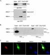
NS1 and Ubc9 interact in vitro and colocalize in the nucleus. (A) GST or Ubc9-GST was expressed in E. coli, bound to glutathione beads, and incubated with lysates from HEK293T cells expressing HA-tagged NS1 from A/Duck/Hubei/L-1/2004 (H5N1). Bound proteins were eluted and analyzed by SDS-PAGE and Western blotting with anti-HA or anti-GST antibody. Whole-cell lysates are shown as input. (B) GST pull-down experiments were done as described above, with lysates from 293T cells expressing either the HA-tagged RBD (NS11-73) or the HA-tagged effector domain (NS174-225) of NS1. Bound proteins were eluted and analyzed by SDS-PAGE and Western blotting with anti-HA antibody. (C) Colocalization of H5N1 NS1 and Ubc9. HeLa cells were cotransfected with pCAGGS-NS1 and pEGFP-Ubc9. Cells were fixed and permeabilized. NS1 was stained with mouse anti-HA monoclonal antibody followed by goat anti-mouse-Cy3 (red) as the secondary antibody. Nuclei were stained with DAPI (blue).
FIG. 3.
FIG. 3.
Sumoylation of A/Duck/Hubei/L-1/2004 (H5N1) NS1 protein in vivo. (A) Colocalization of NS1 and SUMO1. HeLa cells were cotransfected with pCAGGS-NS1 and pCAGGS-SUMO1. Cells were fixed and permeabilized. NS1 protein was stained with anti-HA-tag rabbit polyclonal antibody, and SUMO1 was stained with mouse anti-SUMO1 monoclonal antibody. Goat anti-rabbit-Alexa 488 (green) and goat anti-mouse-Cy3 (red) were used as secondary antibodies. Nuclei were stained with DAPI (blue). (B) Sumoylation of H5N1 NS1. HEK293T cells were transfected with expression plasmids for HA-tagged NS1, NS1CPSFm, Phd, and Cer-SUMO1, as indicated. (Left) NS1 and Phd were immunoprecipitated with anti-HA agarose (IP), and precipitated proteins were further analyzed by SDS-PAGE and Western blotting (WB) with anti-HA polyclonal antibody. (Right) The samples were probed again with anti-SUMO1 monoclonal antibody. Western blots with anti-actin or anti-HA antibody from whole-cell lysates are shown as loading controls (Input). (C) HEK293T cells were transfected with H5N1 NS1CPSFm together with Cer-SUMO1 or Cer-SUMO1AA. Cells were lysed, and NS1 was immunoprecipitated with anti-HA agarose. NS1 was detected as described above. Western blots with anti-GFP (WB: Cerulean) or anti-actin antibody for whole-cell lysates are shown as Cer-SUMO1 expression and loading controls (Input). The positions of molecular mass standards are marked on the left.
FIG. 4.
FIG. 4.
Deconjugation of SUMO1 from A/Duck/Hubei/L-1/2004 (H5N1) NS1 is regulated by SENP1. (A) HEK293T cells were transfected with expression plasmids for NS1CPSFm, Cer-SUMO1, SENP1, or SENP1mut, as indicated, and total DNA levels were adjusted by adding vector plasmid. NS1 was immunoprecipitated (IP) with anti-HA agarose, and precipitated proteins were further analyzed by SDS-PAGE and Western blotting (WB) with anti-HA polyclonal antibody. (B) Modification of H5N1 NS1 protein with endogenous SUMO1. HEK293T cells were cotransfected with NS1CPSFm, SENP1, or SENP1mut, as indicated. Immunoprecipitation and detection of NS1 were performed as described above. Western blots with anti-SUMO1 or anti-actin antibody for whole-cell lysates are shown as input. The positions of molecular mass standards are marked on the left.
FIG. 5.
FIG. 5.
Sumoylation of A/Duck/Hubei/L-1/2004 (H5N1) NS1 in infected cells. A549 cells were transfected with the indicated siRNA for 24 h, followed by infection with r-H5N1-NS virus (MOI = 5) or mock infection. At 8 h postinfection, cells were lysed in SDS loading buffer containing NEM, followed by SDS-PAGE and Western blotting with antibodies against NS1 or actin. The positions of molecular mass standards are marked on the left. NC, control siRNA.
FIG. 6.
FIG. 6.
A/Duck/Hubei/L-1/2004 (H5N1) NS1 is sumoylated at lysine 219 and lysine 221. (A) Mapping of lysine residues responsible for sumoylation of H5N1 NS1. HEK293T cells were transfected with expression plasmids encoding H5N1 NS1CPSFm or mutants containing lysine (K)-to-arginine (R) substitutions at the indicated positions. Cer-SUMO1 or Cer-SUMO1AA was cotransfected as shown. Cells were lysed, and NS1 was immunoprecipitated with anti-HA agarose. NS1 was detected in Western blots with anti-HA polyclonal antibody. (B) Wild-type H5N1 NS1 or NS1K219,221R was coexpressed with Cer-SUMO1 or Cer-SUMO1AA, as indicated, and NS1 was visualized as described above. The positions of molecular mass standards are marked on the left.
FIG. 7.
FIG. 7.
Increased stability of wild-type NS1 (from A/Duck/Hubei/L-1/2004 [H5N1]) compared to the sumoylation-deficient NS1K219,221R mutant. (A) SUMO1 conjugation of A/Duck/Hubei/L-1/2004 (H5N1) NS1 protein inhibits reporter gene activity. A firefly luciferase reporter plasmid under the control of the IFN-β promoter (pIFNβ-Luc) and a Renilla luciferase plasmid (PRL-SV40) under the control of a constitutive SV40 promoter (pSV40-Ren) were cotransfected with the indicated NS1-expressing plasmids into HEK293T cells. After 16 h, cells were infected with 10 HA units/well SeV or mock infected. Cell extracts were collected at 16 h postinfection, and the luciferase activities were measured. Results are given as percentages of the luciferase activity in SeV-infected vector-expressing cells (considered 100%) and represent the averages for three independent experiments, with standard deviations. *, statistically significant (P < 0.05) by Student's t test. (B) Cell extracts from panel A were diluted 5 times and further analyzed by Western blotting (WB) with anti-HA or anti-actin antibody. (C) HEK293T cells were cotransfected with pCAGGS-SUMO1 and NS1 or NS1K219,221R, as indicated. Thirty-six hours after transfection, cells were treated with 100 μg/ml CHX for the indicated periods. NS1 proteins were immunoprecipitated with anti-HA agarose (IP: HA), and precipitated proteins were further analyzed by SDS-PAGE and Western blotting with anti-HA polyclonal antibody to detect NS1 proteins (WB: HA). Western blots of whole-cell lysates with anti-actin antibody are shown as a loading control (Input). The positions of molecular mass standards are marked on the left. (D) Western blots as described for panel C were analyzed by densitometry. Means for three independent experiments (+ standard deviations) are shown.
FIG. 8.
FIG. 8.
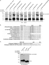
Sumoylation of NS1 proteins from different subtypes and strains. (A) HEK293T cells were cotransfected with plasmids encoding HA-tagged NS1 proteins from different influenza A virus subtypes and strains together with Cer-SUMO1 or Cer-SUMO1AA. Cells were lysed, and NS1 was immunoprecipitated (IP) with anti-HA agarose. Precipitated proteins were further analyzed by SDS-PAGE and Western blotting (WB) with anti-HA polyclonal antibody. The positions of molecular mass standards are marked on the left. (B) Schematic representation of the C-terminal amino acid sequences of NS1 proteins from different influenza A virus strains. Lysine (K) residues (shaded) indicate SUMO1 modification sites for each NS1 protein. (C) (Top) Schematic representation of the C-terminal amino acid sequences of swNS1 and swNS1STOPmut. (Bottom) Swine-origin NS1 (swNS1) and its extended form (swNS1STOPmut) are not sumoylated. HEK293T cells were transfected with H5N1 NS1 (from A/Duck/Hubei/L-1/2004) or with swNS1 (from A/Sichuan/1/2009 [H1N1]) or its C-terminally extended form, swNS1STOPmut, together with Cer-SUMO1 or Cer-SUMO1AA, as indicated. Cells were lysed, and NS1 was immunoprecipitated with anti-HA agarose. NS1 was detected with anti-HA antibody. The positions of molecular mass standards are marked on the left.
FIG. 9.
FIG. 9.
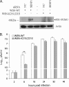
Sumoylation of K219 and K221 of NS1 accelerates virus replication. (A) A549 cells were transfected with the indicated siRNA for 24 h, followed by infection with wild-type WSN-WT (A/WSN/33) or a recombinant WSN virus expressing the NS1K219,221E mutant protein (WSN-K219,221E) at an MOI of 5. At 8 h postinfection, cells were lysed in SDS loading buffer containing NEM, followed by SDS-PAGE and Western blotting with anti-NS1 or anti-actin antibody. The positions of molecular mass standards are marked on the left. (B) A549 cells were infected with either WSN-WT or WSN-K219,221E virus at an MOI of 0.1, and virus production at the indicated times postinfection was determined by plaque assay on MDCK cells. Means for three independent experiments (+ standard deviations) are shown. **, P < 0.01.

Similar articles

Cited by

References

    1. Bailey, D., and P. O'Hare. 2002. Herpes simplex virus 1 ICP0 co-localizes with a SUMO-specific protease. J. Gen. Virol. 83:2951-2964. - PubMed
    1. Basler, C. F., A. H. Reid, J. K. Dybing, T. A. Janczewski, T. G. Fanning, H. Zheng, M. Salvatore, M. L. Perdue, D. E. Swayne, A. Garcia-Sastre, P. Palese, and J. K. Taubenberger. 2001. Sequence of the 1918 pandemic influenza virus nonstructural gene (NS) segment and characterization of recombinant viruses bearing the 1918 NS genes. Proc. Natl. Acad. Sci. U. S. A. 98:2746-2751. - PMC - PubMed
    1. Bergmann, M., A. Garcia-Sastre, E. Carnero, H. Pehamberger, K. Wolff, P. Palese, and T. Muster. 2000. Influenza virus NS1 protein counteracts PKR-mediated inhibition of replication. J. Virol. 74:6203-6206. - PMC - PubMed
    1. Boggio, R., and S. Chiocca. 2006. Viruses and sumoylation: recent highlights. Curr. Opin. Microbiol. 9:430-436. - PMC - PubMed
    1. Boggio, R., R. Colombo, R. T. Hay, G. F. Draetta, and S. Chiocca. 2004. A mechanism for inhibiting the SUMO pathway. Mol. Cell 16:549-561. - PubMed

Publication types

MeSH terms

  NODES
twitter 2