. 2010 Nov 13:11:83.
doi: 10.1186/1471-2199-11-83.

Identification of amino acid residues in protein SRP72 required for binding to a kinked 5e motif of the human signal recognition particle RNA

Affiliations

Identification of amino acid residues in protein SRP72 required for binding to a kinked 5e motif of the human signal recognition particle RNA

Elena Iakhiaeva et al. BMC Mol Biol. .

Abstract

Background: Human cells depend critically on the signal recognition particle (SRP) for the sorting and delivery of their proteins. The SRP is a ribonucleoprotein complex which binds to signal sequences of secretory polypeptides as they emerge from the ribosome. Among the six proteins of the eukaryotic SRP, the largest protein, SRP72, is essential for protein _targeting and possesses a poorly characterized RNA binding domain.

Results: We delineated the minimal region of SRP72 capable of forming a stable complex with an SRP RNA fragment. The region encompassed residues 545 to 585 of the full-length human SRP72 and contained a lysine-rich cluster (KKKKKKKKGK) at postions 552 to 561 as well as a conserved Pfam motif with the sequence PDPXRWLPXXER at positions 572 to 583. We demonstrated by site-directed mutagenesis that both regions participated in the formation of a complex with the RNA. In agreement with biochemical data and results from chymotryptic digestion experiments, molecular modeling of SRP72 implied that the invariant W577 was located inside the predicted structure of an RNA binding domain. The 11-nucleotide 5e motif contained within the SRP RNA fragment was shown by comparative electrophoresis on native polyacrylamide gels to conform to an RNA kink-turn. The model of the complex suggested that the conserved A240 of the K-turn, previously identified as being essential for the binding to SRP72, could protrude into a groove of the SRP72 RNA binding domain, similar but not identical to how other K-turn recognizing proteins interact with RNA.

Conclusions: The results from the presented experiments provided insights into the molecular details of a functionally important and structurally interesting RNA-protein interaction. A model for how a ligand binding pocket of SRP72 can accommodate a new RNA K-turn in the 5e region of the eukaryotic SRP RNA is proposed.


Figures

Figure 1
Figure 1
Participants in the interaction between protein SRP72 and the SRP RNA. a. Wildtype and mutant human SRP72 polypeptide fragments used for investigating their potential to interact with the 5e SRP RNA. Their names are given on the left. Amino acid residue numbering is according to the full-length human SRP72. Indicated are the mutated residues (underlined) and their effects on RNA-binding are shown by the colored dots. Green is for binding activities similar to the 72 h or 72 k fragments, red for no or weak binding, and pink for intermediate activities. The gray circle indicates that mutant polypeptide 72 i was prone to degradation. Potential cutting sites for Chymotrypsin (CT) are shown on top. The RNA binding region with its PDPXRWLPXXER motif (boxed) is indicated by the green brace. b. Outline of the human SRP. The RNA helices in the secondary structure (gray) are numbered from two to eight. The 5' and 3' ends and the two SRP domains are indicated, and the SRP proteins (circles and ovals) are positioned accordingly. The location of the 5e motif is shown in green as are the residue positions 100 and 240 of a neighboring loop. c. Predicted secondary structure of the 5e RNA. Numbering is according to full-length human SRP RNA excluding the helix-closing tetraloop (gray). The 11-nucleotide 5e motif is shown boxed.
Figure 2
Figure 2
5e RNA binding activities of polypeptides 72 e, 72 f, 72 g, 72 h and 72 j. Purified his-tagged polypeptides 72 e to 72 j were incubated with in vitro transcribed 5e SRP RNA and Ni-NTA magnetic agarose beads as described in the Methods. The bound protein and RNA were analyzed by SDS PAGE followed by staining of the same gel with Coomassie blue (lanes labeled p) and Ethidium bromide (lanes labeled r). Molecular mass markers in kDa are shown in lane m. Plus signs indicate the formation of complexes, the minus sign below 72 g indicates that this polypeptide was unable to bind. Variable amounts of a material which probably represented 5e SRP RNA dimers were observed (arrow heads).
Figure 3
Figure 3
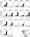
5e SRP RNA binding activities of mutated SRP72 fragments. For each construct, 82.5 ng of in vitro transcribed 5e SRP RNA was incubated with purified protein at the concentrations 0.056, 0.18, 0.56, and 1.8 uM. Samples were incubated with Ni-NTA magnetic agarose beads as described in the Methods. The bound protein and RNA were analyzed by SDS PAGE followed by staining with Coomassie blue (labeled p below each panel) and Ethidium bromide. The amount of bound RNA was measured and normalized to 100 percent in reference to the binding observed with 72 h, 72 j, or 72 k. The insert on the bottom right depicts the design of the assay.
Figure 4
Figure 4
Chymotryptic protection in the RNA binding domain of SRP72. a. Digestion of purified 72 j (lane 1) with Chymotrypsin for 3, 6 or 10 minutes (lanes 2 to 4). Similarly, digestion of purified 72 k (lane 5) for 3, 6 or 10 minutes (lanes 6 to 8). Lane 9, the 72 l fragment (lacking residues 584 to 590) treated with Chymotrypsin for 10 minutes; lane 10, the 585/6 polypeptide, generated by a 10 minute chymotryptic digest of 72 k, retained on Ni-NTA magnetic agarose beads (see Methods for details). All samples were analyzed by electrophoresis on 12.5 percent SDS polyacrylamide gels followed by staining of the polypeptides with Coomassie blue. Molecular mass markers in kDa are indicated on the left and the migrations of Chymotrypsin (CT) and the his-tagged C-terminal 585/6 fragment are marked on the right. b. Digestion of 1.2 μg 72 j with Chymotrypsin in the absence of RNA (lane 1) or in the presence of increasing amounts of human Δ35 RNA (lanes 2 to 4)[23]. Indicated are molecular mass markers and the migrations of Chymotrypsin (CT), the 72 j polypeptide, and the his-tagged oligopeptide generated by the cleavages of the Tyrosines 585 or 586. c. Binding of human Δ35 to increasing amounts of 72 j. d. Results from gelshift experiment for the binding data shown in Figure 5c. Indicated are the mobilities of the free Δ35 RNA (fr) and its complex with 72 j (cp). Lane numbers correspond to the seven data points shown in Figure 5c. e. Similar to Figure 5 d, showing the formation of a complex between the 72 j and the 5e SRP RNA.
Figure 5
Figure 5
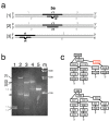
Analysis of 5e SRP RNA kinking. a. Polynucleotide fragment assemblies to investigate 5e SRP RNA kinking. In assembly 1, the 5e motif was placed in the middle, residues 240-AUC-242 were deleted in assembly 2, and the 5e motif was placed near the end in assembly 3. Gray and black lines indicate DNA and RNA, respectively. The sequences of the fragments are provided as Additional File 1. b. The annealed polynucleotide fragments were separated by electrophoresis on an eight percent native polyacrylamide gel followed by staining with Ethidium bromide as described in Methods. Lane 1, annealed fragments 1 and 2 (1-2) and annealed fragments 3 and 4 (3-4); lane 2, same as lane 2 with added r1 and r2; lane 3, same as lane 1 with added r1 and r3; lane 4, annealed fragments 5, 6, r1 and r4; lane 5, annealed fragments 5 and 6; lane m, 100 base pair DNA ladder as indicated. The migration distances of the three assemblies, the annealed fragments and some intermediates (i) are labeled on the left. c. Base arrangements in the predicted 5e SRP RNA K-turn (top) in comparison with the Kt-7 kink-turn (bottom)[27].
Figure 6
Figure 6
Three-dimensional model of the 5e RNA binding fragment of human SRP72. a. Ribbon representation of the three-dimensionaI model of the complete human SRP72 as predicted by I-TASSER [29] and modified as described in Methods. The N- and C-termini are indicated and the atoms of the residues in RNA binding region (532 to 590) are shown as spheres and colored by charge. b. Closeup of the SRP72 RNA binding domain with the identified minimal region required for forming a complex with the 5e SRP RNA in a surface representation. The conserved tryptophan residue located inside the model is drawn in more detail. Shown are the locations of some lysine residues, the prominent chymotryptic cleavage site (CT585/6) and the tyrosine at position 566. c. Same as panel b but viewed at an approximately 90 degree rotation around the y axis revealing a pocket suitable for binding to the kinked 5e SRP RNA (red).

Similar articles

Cited by

References

    1. Stengel KF, Holdermann I, Cain P, Robinson C, Wild K, Sinning I. Structural basis for specific substrate recognition by the chloroplast signal recognition particle protein cpSRP43. Science. 2008;321:253–256. doi: 10.1126/science.1158640. - DOI - PubMed
    1. Koch HG, Moser M, Muller M. Signal recognition particle-dependent protein _targeting, universal to all kingdoms of life. Rev Physiol Biochem Pharmacol. 2003;146:55–94. full_text. - PubMed
    1. Nagai K, Oubridge C, Kuglstatter A, Menichelli E, Isel C, Jovine L. Structure, function and evolution of the signal recognition particle. EMBO J. 2003;22:3479–3485. doi: 10.1093/emboj/cdg337. - DOI - PMC - PubMed
    1. Doudna JA, Batey RT. Structural Insights into the Signal Recognition Particle. Annu Rev Biochem. 2004;73:539–557. doi: 10.1146/annurev.biochem.73.011303.074048. - DOI - PubMed
    1. Halic M, Beckmann R. The signal recognition particle and its interactions during protein _targeting. Curr Opin Struct Biol. 2005;15:116–125. doi: 10.1016/j.sbi.2005.01.013. - DOI - PubMed

Publication types

LinkOut - more resources

  NODES
twitter 2