Comparative Study
. 2011 Jun 15;183(12):1625-32.
doi: 10.1164/rccm.201009-1452OC. Epub 2011 Mar 25.

Airway fibroblasts in asthma manifest an invasive phenotype

Affiliations
Comparative Study

Airway fibroblasts in asthma manifest an invasive phenotype

Jennifer L Ingram et al. Am J Respir Crit Care Med. .

Abstract

Rationale: Invasive cell phenotypes have been demonstrated in malignant transformation, but not in other diseases, such as asthma. Cellular invasiveness is thought to be mediated by transforming growth factor (TGF)-β1 and matrix metalloproteinases (MMPs). IL-13 is a key T(H)2 cytokine that directs many features of airway remodeling through TGF-β1 and MMPs.

Objectives: We hypothesized that, in human asthma, IL-13 stimulates increased airway fibroblast invasiveness via TGF-β1 and MMPs in asthma compared with normal controls.

Methods: Fibroblasts were cultured from endobronchial biopsies in 20 subjects with mild asthma (FEV(1): 90 ± 3.6% pred) and 17 normal control subjects (FEV(1): 102 ± 2.9% pred) who underwent bronchoscopy. Airway fibroblast invasiveness was investigated using Matrigel chambers. IL-13 or IL-13 with TGF-β1 neutralizing antibody or pan-MMP inhibitor (GM6001) was added to the lower chamber as a chemoattractant. Flow cytometry and immunohistochemistry were performed in a subset of subjects to evaluate IL-13 receptor levels.

Measurements and main results: IL-13 significantly stimulated invasion in asthmatic airway fibroblasts, compared with normal control subjects. Inhibitors of both TGF-β1 and MMPs blocked IL-13-induced invasion in asthma, but had no effect in normal control subjects. At baseline, in airway tissue, IL-13 receptors were expressed in significantly higher levels in asthma, compared with normal control subjects. In airway fibroblasts, baseline IL-13Rα2 was reduced in asthma compared with normal control subjects.

Conclusions: IL-13 potentiates airway fibroblast invasion through a mechanism involving TGF-β1 and MMPs. IL-13 receptor subunits are differentially expressed in asthma. These effects may result in IL-13-directed airway remodeling in asthma.


Figures

Figure 1.
Figure 1.
Photomicrographs indicating airway fibroblast invasion through the Matrigel membrane. (A) Image of airway fibroblasts isolated from a representative subject with asthma invading Matrigel toward serum-free media alone as untreated control. (B) Image of airway fibroblasts isolated from the same representative subject with asthma as A invading Matrigel toward IL-13 (50 ng/ml) in serum-free media. (C) Image of airway fibroblasts isolated from a representative normal control subject invading Matrigel toward serum-free media alone as untreated control. (D) Image of airway fibroblasts isolated from the same representative normal control subject as C invading Matrigel toward IL-13 (50 ng/ml) in serum-free media.
Figure 2.
Figure 2.
IL-13 stimulates airway fibroblast invasion of Matrigel in asthma. (A) Airway fibroblast invasiveness at baseline. No significant difference in invasion between groups was observed for the unstimulated airway fibroblasts (P = 0.30). (B) Airway fibroblasts were investigated in the Matrigel assay with 50 ng/ml IL-13 or serum-free media (as a negative control) in the lower chamber of the transwell for a 48-hour incubation period. IL-13 exerts a significant increase in mean numbers of invading fibroblasts in asthma compared with unstimulated control (P = 0.04); however, no effect of IL-13 was observed within the normal control subjects (P = 0.59). (C) IL-13 induces a significant increase in airway fibroblast invasion in asthma. Compared with the normal control subjects, IL-13 significantly induced airway fibroblast invasion in asthma (P = 0.005). Data are expressed as fold change in the mean numbers of airway fibroblasts invading the Matrigel as normalized to the negative control. Subjects with asthma (n = 20; FEV1: 90 ± 3.6%) and normal control subjects (n = 17; FEV1: 102 ± 2.9%), mean ± SEM.
Figure 3.
Figure 3.
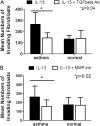
IL-13 stimulates airway fibroblast invasion in asthma. (A) Airway fibroblasts were investigated in the Matrigel assay with a range of concentrations of IL-13 (5–100 ng/ml) or serum-free media (0 ng/ml) in the lower chamber of the transwell for a 48-hour incubation period. The 50 ng/ml concentration of IL-13 induced significantly increased invasion in asthma compared with normal control fibroblasts (solid diamond: P = 0.04). Both the 50 ng/ml (#) and the 100 ng/ml (*) concentrations of IL-13 induced significantly increased numbers of invading cells in asthma compared with the 0, 5, and 10 ng/ml concentrations of IL-13 (P < 0.03). No significant differences in invasion in response to IL-13 were observed for the normal control fibroblasts (P > 0.08 for all comparisons). Subjects with asthma (n = 8; FEV1: 85 ± 5.2%) and normal control subjects (n = 5; FEV1: 105 ± 3%), mean ± SEM. (B) IL-13 neutralizing antibody blocked IL-13–induced airway fibroblast invasion in asthma. Significant reduction in IL-13–induced (50 ng/ml) airway fibroblast invasion was observed in response to 10 μg/ml IL-13 neutralizing antibody in asthma (P = 0.02), but not in the normal control fibroblasts (P = 0.35) or in the unstimulated (negative control) airway fibroblast invasion (P = 0.38) (data not shown). Subjects with asthma (n = 7; FEV1: 78 ± 5.3%) and normal control subjects (n = 5; FEV1: 108 ± 6.4%), mean ± SEM.
Figure 4.
Figure 4.
Transforming growth factor (TGF)-β neutralizing antibody and pan-matrix metalloproteinase (MMP) inhibitor block IL-13–induced airway fibroblast invasion in asthma. (A) Significant reduction in IL-13–induced (50 ng/ml) airway fibroblast invasion was observed in response to 10 μg/ml TGF-β1 neutralizing antibody in asthma (P = 0.04), but not in the normal control fibroblasts (P = 0.15) or in unstimulated (negative control) asthmatic airway fibroblast invasion (P = 0.29) (data not shown). (B) A pan-MMP inhibitor (GM6001; 10 μM) elicited significant reduction in asthmatic airway fibroblast invasion in response to 50 ng/ml IL-13 (P = 0.02); however, no effect was observed in the normal control fibroblasts (P = 0.82). Subjects with asthma (n = 13; FEV1: 92 ± 4.8%) and normal control subjects (n = 9; FEV1: 108 ± 4.0%), mean ± SEM.
Figure 5.
Figure 5.
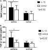
IL-13 significantly stimulates matrix metalloproteinase (MMP)-2 and transforming growth factor (TGF)-β1 secretion by airway fibroblasts in asthma. A subset of airway fibroblasts was grown to confluence in 10% fetal bovine serum and Dulbecco's modified Eagle medium. Cells were rendered quiescent by incubating in serum-free media for 24 hours before incubating with 50 ng/ml IL-13 in serum-free media or serum-free media alone as untreated control for a 24-hour (A, MMP-2) or 48-hour (B, TGF-β1) time course. Cell culture supernatants were collected and acid-treated for measurement of total TGF-β1 or diluted ×5 for measurement of total MMP-2 using colorimetric sandwich ELISA (R&D Systems, Minneapolis, MN). Subjects with asthma (n = 9; FEV1: 88 ± 7.1%) and normal control subjects (n = 6; FEV1: 102 ± 4.9%), mean ± SEM.
Figure 6.
Figure 6.
Airway fibroblast invasion is correlated with methacholine PC20. Single linear regression of log2-transformed methacholine PC20 data from subjects with asthma reveals significant and inverse correlation with mean numbers of invading airway fibroblasts in the Matrigel assay after incubation with 50 ng/ml IL-13 for 48 hours (r = −0.62; P = 0.0005). Subjects with asthma (n = 15, FEV1: 91 ± 4%). Data points are mean numbers of IL-13–induced invading fibroblasts from duplicate or triplicate experiments for each subject.
Figure 7.
Figure 7.
IL-13 receptor subunits expression is significantly augmented in asthmatic tissue. We observed significantly elevated expression of each of the IL-13 receptor subunits (IL-13Rα1, IL-13Rα2, and IL-4Rα) in asthma compared with normal control (P < 0.05). (A) IL-13Rα1, (D) IL-4Rα, and (G) IL-13Rα2 immunohistochemical staining of airway biopsy sections from eight subjects with asthma and eight normal subjects. Data are expressed as mean volume percent positive IL-13Rα1, IL-4Rα, or IL-13Rα2 immunostained cells ± SEM. Representative images of IL-13 receptor subunit immunostaining in normal control (B, E, H) and asthmatic (C, F, I) airway biopsies. Note positive staining in epithelial cells, inflammatory cells, and smooth muscle bundles (arrows).
Figure 8.
Figure 8.
Cell surface receptor expression of IL-13Rα2, IL-13Rα1, and IL-4Rα on airway fibroblasts as measured by flow cytometry. Median fluorescence intensity ± SEM of each of the IL-13 receptor subunits was evaluated using flow cytometry in airway fibroblasts from eight subjects with asthma (FEV1: 83 ± 5.1%) and six normal subjects (FEV1: 104 ± 3.7%). At baseline, airway fibroblast cell surface expression of IL-13Rα2 is significantly reduced in asthma compared with normal control (P = 0.04), whereas expression of IL-13Rα1 and IL-4Rα is not significantly different between subject groups.

Similar articles

Cited by

References

    1. Zhu Z, Homer RJ, Wang Z, Chen Q, Geba GP, Wang J, Zhang Y, Elias JA. Pulmonary expression of interleukin-13 causes inflammation, mucus hypersecretion, subepithelial fibrosis, physiologic abnormalities, and eotaxin production. J Clin Invest 1999;103:779–788. - PMC - PubMed
    1. Lee CG, Homer RJ, Zhu Z, Lanone S, Wang X, Koteliansky V, Shipley JM, Gotwals P, Noble P, Chen Q, et al. Interleukin-13 induces tissue fibrosis by selectively stimulating and activating transforming growth factor beta(1). J Exp Med 2001;194:809–821. - PMC - PubMed
    1. Ingram JL, Antao-Menezes A, Mangum JB, Lyght O, Lee PJ, Elias JA, Bonner JC. Opposing actions of Stat1 and Stat6 on IL-13-induced up-regulation of early growth response-1 and platelet-derived growth factor ligands in pulmonary fibroblasts. J Immunol 2006;177:4141–4148. - PubMed
    1. Kraft M, Lewis C, Pham D, Chu HW. IL-4, IL-13, and dexamethasone augment fibroblast proliferation in asthma. J Allergy Clin Immunol 2001;107:602–606. - PubMed
    1. LaPorte SL, Juo ZS, Vaclavikova J, Colf LA, Qi X, Heller NM, Keegan AD, Garcia KC. Molecular and structural basis of cytokine receptor pleiotropy in the interleukin-4/13 system. Cell 2008;132:259–272. - PMC - PubMed

Publication types

Substances

  NODES
Note 1
twitter 2