Review
. 2011 Apr;91(2):691-731.
doi: 10.1152/physrev.00004.2010.

Tissue organization by cadherin adhesion molecules: dynamic molecular and cellular mechanisms of morphogenetic regulation

Affiliations
Review

Tissue organization by cadherin adhesion molecules: dynamic molecular and cellular mechanisms of morphogenetic regulation

Carien M Niessen et al. Physiol Rev. 2011 Apr.

Abstract

This review addresses the cellular and molecular mechanisms of cadherin-based tissue morphogenesis. Tissue physiology is profoundly influenced by the distinctive organizations of cells in organs and tissues. In metazoa, adhesion receptors of the classical cadherin family play important roles in establishing and maintaining such tissue organization. Indeed, it is apparent that cadherins participate in a range of morphogenetic events that range from support of tissue integrity to dynamic cellular rearrangements. A comprehensive understanding of cadherin-based morphogenesis must then define the molecular and cellular mechanisms that support these distinct cadherin biologies. Here we focus on four key mechanistic elements: the molecular basis for adhesion through cadherin ectodomains, the regulation of cadherin expression at the cell surface, cooperation between cadherins and the actin cytoskeleton, and regulation by cell signaling. We discuss current progress and outline issues for further research in these fields.


Figures
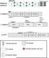
Figure 1
Figure 1. Domain structure of the cadherin-catenin complex components
Schematic overview of the domain structure of a classical cadherin and its three associated catenins, β-catenin, α-catenin and p120-ctn.
Figure 2
Figure 2. The cadherin-catenin complex
(A) Schematic representation of classical cadherins in C. elegans, Drosophila and vertebrates. (B) Schematic representation of the vertebrate cadherin-catenin complex. (C) Structural model of the cadherin catenin complex. This model is based on the crystal structures of the C-cadherin extracellular domain, the cadherin cytoplasmic domain bound to the armadillo repeats of β-catenin, the cadherin juxtamembrane domain bound to the p120-4AΔins and a-catenin fragments. Model reproduced with permission from (152).
Figure 3
Figure 3. Physiological role for cadherins in tissue integrity
(A) Cadherin function in stratifying epithelium of the skin. Loss of all classical cadherins (E- and P-cadherin) in this tissue results in loosening of the intercellular contacts associated with loss of epidermal barrier function. (B) Loss of cadherin expression in epithelia results in loosening of contacts and the acquisition of more migratory behavior. This is a key feature of cells that undergo epithelial/mesenchymal transition. Vice versa, E-cadherin expression is induced during mesenchymal-epithelial transition resulting increased cellular adhesion and an epithelial appearance. EMT and MET are important processes not only during morphogenesis but also contribute to carcinogenesis. (C) Synapse formation. Loss of cadherins affects neuronal transmission and connectivity. Cadherin adhesive interactions are crucial for proper synapse formation between neurons as well as the neuro-muscular junctions. (D) Cadherins are required for the establishment of stable adherens junctions during morphogenetic movements. In C. elegans during ventral closure the embryonic epidermis spreads from the dorsal side and the leading cells of the two free edges meet at the ventral midline to enclose the embryo (left side). The leading edge cells extend filopodial protrusions that rapidly establish stable adherens junctions upon contact with an opposite filopodia, thereby rapidly increasing the contact sites between opposite cells resulting in sealing of the sheet. In the absence of the cadherin/catenin complex embryos are unable to form adherens junctions between opposite leading cells and thus cannot enclose the embryo.
Figure 4
Figure 4. The role of cadherin in cell sorting and positioning
(A) Differential type or levels of cadherin expression on cells drive cell sorting in vitro in cell (re)aggregation assays. (B) During the formation of the neural tube E-cadherin is switched off in a subset of ectodermal cells, whereas N-cadherin expression is turned on in these cells (red cell membranes) driving segregation of these cell populations. Other in vivo examples are neural crest cell migration and positioning and segregation of motor neuron cells. (C) Drosophila oocyte positioning where differential cadherin expression in the follicle cells is crucial to properly position the oocyte at the posterior end of the embryo. For detailed description see text.
Figure 5
Figure 5. Cadherins and cell shape: illustration of the cell organization in the ommatidium of the Drosophila retina
Each ommatidium in the compound eye comprises 20 cells. At the center are two anterior/posterior cone cells (C1) and two equatorial cone cells (C2). The cone cells are enclosed by two primary pigment cells P1. All cell boundaries express E-cadherin, but the boundaries between the cone cells express both N- and E-cadherin. The red indicates a genotypic marker for N-cadherin. The upper left panel shows the cell organization in a normal fly, and the lower panel is the cell organization predicted by a simple mechanical model that considered only adhesion energies and membrane elasticity. The right panels show the cell organization in a mutant fly in which the left cone cell (black) indicated by the red lines (lower panel) lacks N-cadherin. The effect of this deletion is accurately predicted by the mechanical model. (Adapted from (136))
Figure 6
Figure 6. Cadherin in morphogenetic movements
Cadherin function is essential for morphogenetic movements, such as cell-on-cell motility, epiboly, convergent extension movements or migration of intestinal epithelial cells along the crypt/villi axis. (A) Convergence extension movements in which cells align in the plane of the tissue and intercalate to e.g. drive anterior-posterior extension of tissues and/or embryos. Cadherins are crucial to establish contacts and through local modulation of the cytoskeleton provide the pulling force for intercalation. Either overexpression or loss of cadherins interferes with convergent extension movements in e.g. Xenopus or Zebrafish. (B) Drosophila border cells that through a complex signaling pathway switch on E-cadherin to migrate on E-cadherin expressing nurse cells. Loss of E-cadherin on either nurse or border cells prevents migration (see text for more details).
Figure 7
Figure 7. Role of the EC1 domain in cell sorting in vitro
(A) Cells (red and blue circles) expressing E- or P-cadherin (red and blue ectodomains, respectively) sort out when mixed together. (B) If the EC1 domain of P-cadherin (blue) was replaced with the EC1 domain of E-cadherin (red), then cells expressing the E/P-cadherin chimera (blue) intermixed with cells expressing E-cadherin (red).
Figure 8
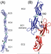
Figure 8. Classical Cadherin Structure and Model of Homophilic Binding
(A) Crystal structure of the extracellular region (EC1-5) of Xenopus C-cadherin. (B) Binding between N-terminal domains of the C-cadherin extracellular region in which the Trp2 (W2) residues from opposing cadherins dock into the hydrophobic pocket of the opposed protein. Reproduced with permission from (199).
Figure 9
Figure 9. Possible mechanism(s) of cadherin binding consistent with biophysical and structural data
(A) Putative cis (lateral) bonds, possibly mediated by EC3 domains (grey ovals) stabilize lateral cadherin dimers. (B) The rapid, initial formation of trans bonds between EC1 domains (white ovals) facilitates cadherin accumulation at cell-cell junctions, and the subsequent slower lateral oligomerization via EC3 domains. (C) Cadherin flexibility could also enable the formation of EC3-dependent between cadherins on opposing membranes or between relatively unconstrained, opposing cadherins in force probe measurements. In (D) EC3 domains bind in an anti-parallel alignment between opposed membranes.
Figure 10
Figure 10. Surface organization of cadherins
Monomers, dimers and oligomers of varying size are found on the cell surface and likely exist in dynamic equilibrium with one another. Adhesive ligation promotes oligomers and clusters, a process that also requires cytoplasmic factors including p120-ctn, signaling molecules, and elements of the acto-myosin cytoskeleton.
Figure 11
Figure 11. Itinerary of cadherins within the cell
Newly synthesized cadherins are transported from the Golgi apparatus (GA) to the cell surface (1), where they are available to engage in cis-binding interactions (2) and form higher-order oligomers and clusters (3). Alternatively, free cadherin, that unable to engage in cis-interactions or which unbinds, is endocytosed (4). Following internalization (5), cadherins are trafficked to early sorting endosomes (EE/SE) from which they may transported back to the cell surface via Rab11-positive recycling endosomes (RE, 6,7), or trafficked through late endosomes/multivesciular bodies (LE,8) for degradation in lysosomes (LY, 9). For clarity, only β-catenin and 120-catenin are drawn in this diagram.
Figure 12
Figure 12. Regulation of cadherin endocytosis in control of surface expression
Surface cadherins can be fated either for stabilization on the surface (1) or for endocytic uptake (2). (1) Stabilization, so that cadherins are not internalized, is promoted by cadherin ligation, masking of dileucine (LL) and tyrosine (Y) residues by p120-ctn, Rac/Cdc42 signaling and the actin cytoskeleton, involving proteins such as IQGAP. (2) Cadherin internalization can be promoted by multiple mechanisms: a) In endothelia, VEGF signaling induces phosphorylation of VE-cadherin ser 665 by a p21-activated kinase (PAK). This promotes binding of b-arrestin and _targeting for endocytosis. b) Alternatively, displacement of p120-catenin unmasks the key dileucine and tyrosine residues that promote clathrin-coated uptake and Hakai binding, respectively.
Figure 13
Figure 13. Consequences of cadherin proteolytic cleavage
Ectodomain shedding by metalloproteases results in the release of the cadherin extracellular domain. This may result in reduced intercellular adhesion by directly reducing the number of functional adhesive complexes at the cell surface. In addition, the released cadherin extracellular domain may also interfere with adhesion. In addition, the extracellular domain can alter local signaling induced by the full length cadherin (e.g. aPKC activation), perhaps by altering clustering or local concentration of cadherins. Upon release of the cadherin extracellular domain, the cytoplasmic domain is subjected to cleavage by presenilins and caspase resulting in release from the membrane and translocation into the nucleus. Here it may modulate either directly or indirectly β-catenin/TCF dependent as well as p120/Kaiso dependent regulation of transcription. The function of the released cytoplasmic domain itself or of a-catenin in the nucleus is at present unclear.
Figure 14
Figure 14. Regulation of actin filament dynamics by cadherin adhesion complexes
Cadherin adhesions may promote actin filament assembly by recruitment of actin nucleators, such as formins and the Arp2/3 complex. Filament growth following nucleation is regulated by other proteins, such as the Ena/VASP proteins, that promote growth of filaments at their barbed ends.
Figure 15
Figure 15. Anchorage of cadherin adhesion complexes to the actin cytoskeleton
Potential models include: A) Binding to cortical actin filaments directly via α-catenin. Although direct binding of cadherin-bound α-catenin to F-actin has not been confirmed in vitro conformational change induced by e.g. mechanical force may allow the cadherin-bound α-catenin to interact with actin filaments. B. Alternative mechanisms to couple cadherin complexes to actin filaments. Potential other mechanisms to physically couple cadherin complexes to F-actin include binding proteins, such as EPLIN, which are recruited by α-catenin. Alternatively, but not exclusively, other actin binding proteins, such as Myosin VI, can interact with cadherins by as-yet-uncharacterized molecular mechanisms.
Figure 16
Figure 16. Models for cadherin-dependent cell signaling
A) Direct cadherin-activated signaling: here adhesive ligation of the cadherin stimulates intracellular signal transduction processes, such as the small GTPase Rac, a process that involves intermediary Guanine nucleotide exchange factors (GEF) such as Tiam-1. B) Juxtacrine signaling: here cadherin adhesion brings cell surfaces together, thereby allowing other contact-dependent signaling mechanisms (e.g. gap junction-mediated cell-cell communication) to be active.
Figure 17
Figure 17. Potential mechanisms for regulation of cadherin biology by tyrosine kinase signaling
A) Uncoupling: tyrosine phosphorylation of the cadherin cytoplasmic tail and/or associated proteins dissociates catenins leading to disassembly of the cadherin molecular complex. B) Scaffolding signaling complexes: tyrosine phosphorylation generates binding sites for association of signaling molecules such as C-terminal kinase (CSK, to VE-cadherin cytoplasmic tail) or PTP1B (to β-catenin). C) Cadherin-based signaling: recruitment of tyrosine kinases as part of a cadherin signaling pathway mediates control of downstream effectors, such as the cytoskeletal regulator, cortactin.

Similar articles

Cited by

References

    1. Abe K, Chisaka O, van Roy F, Takeichi M. Stability of dendritic spines and synaptic contacts is controlled by aN-catenin. Nat Neurosci. 2004 - PubMed
    1. Abe K, Takeichi M. EPLIN mediates linkage of the cadherin catenin complex to F-actin and stabilizes the circumferential actin belt. Proc Natl Acad Sci U S A. 2008;105:13–19. - PMC - PubMed
    1. Adam AP, Sharenko AL, Pumiglia K, Vincent PA. Src-induced tyrosine phosphorylation of VE-cadherin is not sufficient to decrease barrier function of endothelial monolayers. J Biol Chem. 2010;285:7045–7055. - PMC - PubMed
    1. Adams CL, Chen Y-T, Smith SJ, Nelson WJ. Mechanisms of epithelial cell-cell adhesion and cell compaction revealed by high-resolution tracking of E-cadherin-green fluorescent protein. J Cell Biol. 1998;142:1105–1119. - PMC - PubMed
    1. Akhmanova A, Stehbens SJ, Yap AS. Touch, grasp, deliver and control: functional cross-talk between microtubules and cell adhesions. Traffic. 2009;10:268–274. - PubMed

Publication types

LinkOut - more resources

  NODES
Association 1
INTERN 3
todo 4
twitter 2