. 2011 Dec 9;286(49):42690-42703.
doi: 10.1074/jbc.M111.290189. Epub 2011 Oct 3.

Alternative splicing produces Nanog protein variants with different capacities for self-renewal and pluripotency in embryonic stem cells

Affiliations

Alternative splicing produces Nanog protein variants with different capacities for self-renewal and pluripotency in embryonic stem cells

Satyabrata Das et al. J Biol Chem. .

Abstract

Embryonic stem (ES) cells are distinguished by their ability to undergo unlimited self-renewal although retaining pluripotency, the capacity to specify cells of all germ layers. Alternative splicing contributes to these biological processes by vastly increasing the protein coding repertoire, enabling genes to code for novel variants that may confer different biological functions. The homeodomain transcription factor Nanog acts collaboratively with core factors Oct4 and Sox2 to govern the maintenance of pluripotency. We have discovered that Nanog is regulated by alternative splicing. Two novel exons and six subexons have been identified that extend the known Nanog gene structure and protein coding capacity. Alternative splicing results in two novel Nanog protein variants with attenuated capacities for self-renewal and pluripotency in ES cells. Our previous results have implicated the C-terminal domain, including the tryptophan-rich (WR) domain of Nanog, to be important for the function of Nanog (Wang, J., Levasseur, D. N., and Orkin, S. H. (2008) Proc. Natl. Acad. Sci. U.S.A. 105, 6326-6331). Using point mutation analyses, serine 2 (Ser-2) of Nanog has been identified as critical for ES cell self-renewal and for stabilizing a pluripotent gene signature. An inducible conditional knock-out was created to test the ability of new Nanog variants to genetically complement Nanog null ES cells. These results reveal for the first time an expanded Nanog protein coding capacity. We further reveal that a short region of the N-terminal domain and a single phosphorylatable Ser-2 is essential for the maintenance of self-renewal and pluripotency, demonstrating that this region of the protein is highly regulated.


Figures

FIGURE 1.
FIGURE 1.
An extended gene structure of mouse Nanog encodes novel splice isoforms and protein variants. A, schematic of the extended gene and discovered novel variants (left) and the coded proteins (right) of mouse Nanog. The known Nanog a gene structure is depicted at the top. Rectangles (left) designate exons with the shaded sections indicating coding regions. The protein schematic to the right denotes individual Nanog subdomains (ND, N-terminal domain; HD, homeodomain; CD, C-terminal domain; WR, tryptophan-rich domain) with the amino acid numbers indicated above. B, Western blotting of total lysates from the indicated ES cell lines reveals the detection of a ladder of bands with an antibody raised against the N terminus of Nanog (C) qRT-PCR experiments to detect and measure the expression of Nanog transcripts initiating from exon 3A or exon 1B using cytosolic RNA of four different ES cell lines. D, total RNA from mouse blastocysts was analyzed as in C. The data are normalized to Ywhaz and Gapdh, and are graphed with the S.E. of technical replicates.
FIGURE 2.
FIGURE 2.
Recombinant expression of the novel Nanog variants. HEK293T (A) or J1 ES cells (B) were transiently transfected with the cloned V5-His-tagged variants of Nanog, and total lysates were prepared and subjected to Western blotting with anti-V5 antibody. Predicted molecular weight of all expression constructs with the V5-His tag is indicated. Size of protein markers are indicated on the left. Possible ATG usage with each variant is mentioned. The same blot was stripped and subjected to Western blotting with anti-Lamin B antibody to show equivalent loading.
FIGURE 3.
FIGURE 3.
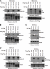
Novel Nanog variants b and c interact with the known isoform Nanog a. A, co-IP in heterologous HEK293T cells. Total lysates were prepared from HEK293T cells transiently transfected with the constructs indicated. Co-IP was performed with anti-V5 antibody (left) or anti-FLAG antibody (right), followed by Western blotting with the antibodies indicated. Interaction of Nanog a with itself (homodimerization) was used as a positive control. Total lysates were subjected to Western blotting with the indicated antibodies as inputs. Nanog b and c are able to interact with Nanog a. B, co-IP in E14T ES cells. The co-IP was repeated with total lysates from transiently transfected E14T cells with the indicated constructs and the interactions of Nanog b and c with Nanog a were verified by Western blotting.
FIGURE 4.
FIGURE 4.
Novel Nanog variants interact with Oct4 and Sall4. A, co-IP of FLAG-tagged Oct4 with V5-His-tagged Nanog isoforms in heterologous HEK293T cells. Total lysates were prepared from HEK293T cells transiently transfected with the constructs indicated. Co-IP was performed with anti-V5 antibody (left) or anti-FLAG antibody (right), followed by Western blotting with the antibodies indicated. Interaction of Oct4 with Nanog a was used as a positive control. Total lysates were subjected to Western blotting with the indicated antibodies as inputs. Nanog b and c are able to interact with Oct4 as well as Nanog a. B, co-IP of FLAG-tagged Oct4 with V5-His-tagged Nanog isoforms in E14T ES cells. Co-IP was repeated with anti-V5 antibody using total lysates from transiently transfected E14T cells with the indicated constructs, and the interactions of Nanog b and c with Oct4 were verified by Western blotting. C, co-IP of endogenous Oct4 in E14T ES cells with V5-His-tagged Nanog isoforms. E14T cells were transiently transfected with the constructs indicated and total lysates were prepared. Co-IP was performed using antibodies directed against V5 (top) or Oct4 (middle), followed by Western blotting with the indicated antibodies. Nontransfected E14T cell total lysates were used as a negative control in the co-IP. Total lysates were subjected to Western blotting with anti-Oct4 as inputs. * indicates nonspecific signal. D, co-IP of FLAG-tagged Sall4 with V5-His-tagged Nanog isoforms in E14T ES cells. Total lysates were prepared from E14T cells transiently transfected with the constructs indicated. Co-IP was performed with anti-V5 antibody (left) or anti-FLAG antibody (right), followed by Western blotting with the antibodies indicated. Interaction of Sall4 with Nanog a was used as a positive control. Total lysates were subjected to Western blotting with the indicated antibodies as inputs. Nanog b and c are able to interact with Sall4 as well as Nanog a.
FIGURE 5.
FIGURE 5.
Nanog a and c, but not Nanog b, sustain LIF-independent self-renewal. Quantitative presentation of the colony formation assay in parental Oct4GiP and stable transfectants expressing the different Nanog variants or their mutants. Cells were plated at clonal density onto gelatin-coated plates and grown in the presence and absence of LIF. After 8 days, colonies were stained for AP and scored into AP positive and negative colonies as indicated on the right. * indicates significantly different numbers of AP positive colonies in comparison to the Oct4GiP control cells grown in the absence of LIF (p < 0.03). The data shown along with the S.E. are from two independent experiments.
FIGURE 6.
FIGURE 6.
Nanog variants enable ES cells to retain self-renewal in the absence of Nanog a. A, loss of Nanog protein in Dox-treated NgcKO cells. Western blot analysis after incubation of the NgcKO cells with 200 ng/ml of Dox for the indicated time periods using anti-Nanog N-terminal antibody. There are undetectable levels of Nanog within 24 h of Dox treatment. For panels A and B, the same blot was stripped and subjected to Western blotting with anti-Lamin B antibody to demonstrate equivalent loading. Relative amounts of Nanog protein are indicated above the blot. B, Western blot analysis of stable clones of different Nanog isoforms expressing (as indicated) NgcKO cells with anti-V5 antibody to detect the V5-tagged transgenes before and after Dox treatment. All clones show similar levels of different Nanog variants. C, V5-tagged Nanog variants were transfected into the NgcKO cells as indicated and stable clones were picked for each of the variants growing in the presence of Dox and subjected to AP staining. All variants enabled morphological rescue of the NgcKO cells to retain typical ES cell morphology and stain positively for AP activity, whereas the untransfected NgcKO cells were differentiated and lost the ability for AP staining in the absence of Nanog after being cultured in the presence of Dox for 5 days (upper row middle panel). Tg, transgene.
FIGURE 7.
FIGURE 7.
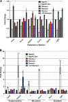
Nanog b and c variants have compromised ability to rescue pluripotency expression markers and to fully repress certain trophectoderal markers. A, qRT-PCR analysis of the expression of specific stem cell pluripotency marker genes in control NgcKO, complete Nanog knockdown, and Nanog variant rescuing NgcKO cells. Stable clones of the Nanog variants in the NgcKO cell line as mentioned were grown in the presence of Dox. Untransfected control NgcKO cells were grown in Dox for 6 days. The data shown are normalized to Gapdh, and shown along with the S.E. of technical replicates. The expression level in the control NgcKO cells is set as 1. B, qRT-PCR analysis of the expression of various cell lineage marker genes in control NgcKO, complete Nanog knockdown, and Nanog variant rescuing NgcKO cells.
FIGURE 8.
FIGURE 8.
Nanog b and c variants do not fully repress primitive endodermal differentiation markers. A, qRT-PCR analysis of the expression of primitive endoderm marker genes in control NgcKO, complete Nanog knockdown, and Nanog variant rescuing NgcKO cells. Stable clones of the Nanog variants in the NgcKO cell line as mentioned were grown in the presence of Dox. Untransfected control NgcKO cells were grown in Dox for 6 days. The data are normalized to Gapdh, and shown along with the S.E. of technical replicates. The expression level in control NgcKO cells is set as 1. B, qRT-PCR analysis of the expression of endodermal marker genes in control NgcKO, complete Nanog knockdown, and Nanog variant rescuing NgcKO cells. C, model of the differential roles for the different Nanog variants. ES cells need to keep the lineage commitment genes suppressed to maintain their self-renewal. The Nanog a variant promotes self-renewal by maintaining expression of the core pluripotency factor network and keeping differentiation promoting transcriptional regulators suppressed. Both the Nanog b and c variants have a reduced capacity to keep primitive endoderm marker genes suppressed. Although Nanog a and b variants are able to sustain Oct4 levels, the c variant does so less efficiently, which can lead to expression of trophectoderm genes. All three variants are able to keep mesodermal, ectodermal, and endodermal markers repressed.

Similar articles

Cited by

References

    1. Evans M. J., Kaufman M. H. (1981) Nature 292, 154–156 - PubMed
    1. Martin G. R. (1981) Proc. Natl. Acad. Sci. U.S.A. 78, 7634–7638 - PMC - PubMed
    1. Orkin S. H., Wang J., Kim J., Chu J., Rao S., Theunissen T. W., Shen X., Levasseur D. N. (2008) Cold Spring Harbor Symp. Quant. Biol. 73, 195–202 - PubMed
    1. Wang J., Rao S., Chu J., Shen X., Levasseur D. N., Theunissen T. W., Orkin S. H. (2006) Nature 444, 364–368 - PubMed
    1. Boyer L. A., Lee T. I., Cole M. F., Johnstone S. E., Levine S. S., Zucker J. P., Guenther M. G., Kumar R. M., Murray H. L., Jenner R. G., Gifford D. K., Melton D. A., Jaenisch R., Young R. A. (2005) Cell 122, 947–956 - PMC - PubMed

Publication types

  NODES
INTERN 4
Note 1
twitter 2