. 2011 Oct 14;147(2):447-58.
doi: 10.1016/j.cell.2011.08.043.

Unidirectional cross-activation of GRPR by MOR1D uncouples itch and analgesia induced by opioids

Affiliations

Unidirectional cross-activation of GRPR by MOR1D uncouples itch and analgesia induced by opioids

Xian-Yu Liu et al. Cell. .

Abstract

Spinal opioid-induced itch, a prevalent side effect of pain management, has been proposed to result from pain inhibition. We now report that the μ-opioid receptor (MOR) isoform MOR1D is essential for morphine-induced scratching (MIS), whereas the isoform MOR1 is required only for morphine-induced analgesia (MIA). MOR1D heterodimerizes with gastrin-releasing peptide receptor (GRPR) in the spinal cord, relaying itch information. We show that morphine triggers internalization of both GRPR and MOR1D, whereas GRP specifically triggers GRPR internalization and morphine-independent scratching. Providing potential insight into opioid-induced itch prevention, we demonstrate that molecular and pharmacologic inhibition of PLCβ3 and IP3R3, downstream effectors of GRPR, specifically block MIS but not MIA. In addition, blocking MOR1D-GRPR association attenuates MIS but not MIA. Together, these data suggest that opioid-induced itch is an active process concomitant with but independent of opioid analgesia, occurring via the unidirectional cross-activation of GRPR signaling by MOR1D heterodimerization.


Figures

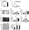
Figure 1
Figure 1. MIS is not Correlated with MIA
(A) Dose effect of i.t. morphine on MIS and MIA in 30 min. (B) Time course of morphine on MIS and MIA. (C) Induction of acute MIA tolerance with morphine (100 mg/kg, s.c.) or saline. Mice had returned to the basal nociceptive latencies 24 hr after injection. (D) Acute MIA tolerance was tested with i.t. morphine 24 hr after s.c. morphine treatment. (E) I.t. morphine induced comparable scratches in acute MIA tolerant and control mice. (F) Induction of chronic MIA tolerance by daily injection of morphine (10 mg/kg, s.c.) and MIA was examined daily. (G) After 5 days of systemic morphine injection, i.t. morphine also showed antinociceptive tolerance. (H) I.t. morphine induced comparable scratches in chronic MIA tolerant and control mice. In all experiments, the dose of i.t. morphine is 0.3 nmol. n = 6~8 per group. MIA results are expressed as % maximum possible effect (MPE). *p < 0.05. Error bars represent standard error of the mean.
Figure 2
Figure 2. Identification of MIA- and MIS-Specific MOR isoforms
(A) MIS was severely impaired in MOR KO mice, whereas GIS in MOR KO mice was comparable to that in wild-type littermate control mice. (B) MIS was significantly reduced by naloxone (3 mg/kg, s.c.). (C) Schematic representation of partial alternative Oprm splicing in the mouse. Clear rectangles represent the _targeting exons by siRNA. (D) MIS was significantly reduced by MOR siRNA _targeting at exon 1 (MOR1, 1C, 1D, and 1E) and exon 9 (MOR1C, 1D, and 1E), but not by siRNA _targeting at exon 4 (MOR1) or exon 7 (MOR1C and 1E). *p < 0.05. Sequence of siRNAs are included in supplementary file. (E) MOR siRNA _targeting at exon 1 and exon 4, but not exon 7 or exon 9 significantly reduced morphine analgesic effect. (F) Representative gel images showing decreased spinal MOR1 mRNA level after exon 1 and exon 4 specific siRNA treatments and decreased spinal MOR1D mRNA level after exon 1 and exon 9 specific siRNA treatments. 18S RNA, an internal control, was comparable among all groups. (G) Exon 1 and exon 4 specific siRNA significantly knocked down MOR1 mRNA in spinal cord as detected by q-RT-PCR. (H) Spinal MOR1D mRNA level was significantly reduced by siRNA specific to MOR exon 1 and exon 9 as detected by qRT-PCR. (I and J) Western blot (I) and quantified data (J) showed MOR exon 9 siRNA treatment specifically reduced protein level of MOR1D but not that of MOR1 or GRPR in the spinal cord. In all experiments, n = 5~8 per group. *p < 0.05. Error bars represent standard error of the mean. Also see Figure S1
Figure 3
Figure 3. Co-Expression of GRPR and MOR1D in Lamina I of the Spinal Cord
(A–C) Double immunostaining of GRPR (red, lamina I) and MOR1D (green, lamina II) revealed no co-localization of GRPR and MOR1 in the dorsal spinal cord. (D–F) Double immunostaining of GRPR (red) and MOR1D (green) in lamina I of the spinal cord. Arrows indicate co-expression (yellow) and arrowheads indicate singular expression. Cells co-expressing GRPR (11/33) and MOR1D (11/17), which represent ~31% of GRPR-positive cells and ~65% of MOR1D-positive cells respectively, were found in 25 lumbar spinal cord sections. (G–I) Double immunostaining revealed no co-localization of GRPR (red, lamina I) and MOR1 (green, lamina II) in the dorsal spinal cord. Scale bar, 50 μm. Also see Figure S2
Figure 4
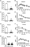
Figure 4. GRPR is Important for Opioid-Induced Scratching Behavior
(A) MIS was nearly abolished in GRPR KO mice compared with wild-type littermate mice. (B) MIA was comparable between GRPR KO and wild-type littermates. (C) Scratching behavior induced by i.t. DAMGO was significantly reduced in GRPR KO mice. (D) Analgesic effect of i.t. DAMGO was comparable between GRPR KO and wild-type littermates. (E and F) Scratching behavior induced by i.t. fentanyl was significantly reduced in GRPR KO mice (E), while the analgesic effect of fentanyl was not affected (F). (G) MIS was significantly inhibited by co-injection with the GRPR antagonist (0.1, 1 nmol). (H) MIA was not significantly affected by co-injection of the GRPR antagonist. In all experiments, the dose of i.t. morphine is 0.3 nmol. n = 6~9 per group. *p < 0.05. Error bars represent standard error of the mean. See also Figure S3.
Figure 5
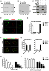
Figure 5. Co-Immunoprecipitation and Co-Internalization of GRPR and MOR1D
(A) Myc-GRPR (43 kDa) was detected in membrane fraction of HA-MOR1/Myc-GRPR cells (L1) and HA-MOR1D/Myc-GRPR cells (L2). Anti-HA antibody co-precipitated Myc-GRPR from HA-MOR1D/Myc-GRPR cells (L4), but not from HA-MOR1/Myc-GRPR cells (L3). (B) Expression of HA-MOR1 (44 kDa) in HA-MOR1/Myc-GRPR cells (L1) and expression of HA-MOR1D (44 kDa) in HA-MOR1D/Myc-GRPR cells (L2) were revealed by anti-HA immunoblotting. An HA-MOR1D band (44 kDa) was precipitated by anti-Myc antibody from HA-MOR1D/Myc-GRPR cells (L4). Anti-Myc antibody failed to precipitate HA-MOR1 from HA-MOR1/Myc-GRPR cells (L3). IP: immunoprecipitaion, IB: immunoblotting, kDa: kilodaltons. (C) GRPR, MOR1D and MOR1 were detected in the membrane extract of dorsal horn (L1). GRPR was co-precipitated by anti-MOR1D (L3) but not by anti-MOR1 (L4) or an non specific rabbit IgG (L2). (D and E) Immunostaining (D) and ELISA (E) revealed internalization of HA-MOR1D but not HA-MOR1 or Myc-GRPR upon morphine treatment, while GRP induced internalization of GRPR but not MOR1D or MOR1. (F and G) Immunostaining (F) and ELISA (G) revealed that Myc-GRPR was co-internalized with MOR1D, but not with MOR1 upon morphine stimulation. GRP only induced internalization of GRPP but not MOR1D or MOR1. (H) Naloxone dose-dependently blocked morphine-induced internalization of Myc-GRPR and HA-MOR1D. (I) The GRPR antagonist blocked morphine-induced internalization of Myc-GRPR, but not HA-MOR1D. Data are expressed as mean and standard error of three independent experiments. Error bars represent standard error of the mean. *p < 0.05. Also see Figure S4.
Figure 6
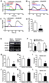
Figure 6. Cross Activation of the GRPR Signal Transduction Pathway by MOR1D in Response to Morphine
The responses of HEK 293 cells expressing vary receptors to morphine or GRP, tested using calcium imaging. (A) HEK 293 cells co-expressing MOR1D and GRPR showed calcium response to both morphine and GRP. Cells co-expressing MOR1 and GRPR were unable to respond to morphine, whereas they responded to GRP. (B) In cells co-expressing MOR1D and GRPR the GRPR antagonist completely blocked morphine- or GRP-induced Ca2+ increase, while naloxone blocked morphine- and reduced GRP-induced Ca2+ response. (C) Both PLC inhibitor U73122 and IP3 receptor antagonist 2-APB blocked the calcium response to morphine and GRP in cells co-expressing MOR1D and GRPR. U73343, an inactive structural analog of U73122 had no such effect. (D) Quantified data comparing peak intracellular calcium concentration. Naloxone significantly reduced GRP-induced [Ca2+]i increase in cells co-expressing MOR1D and GRPR. (E and F) GRPR+ cells in superficial dorsal horn were ablated by bombesin-saporin. The superficial dorsal horn was dissected for qRT-PCR. Gel image (E) and quantitative analysis (F) showed that PLCβ3 mRNA was lost in bombesin-saporin-treated group. PLCβ1 and IP3R3 mRNA were significantly decreased by bombesin-saporin treatment. (G) Two days after the last injection of PLCβ siRNA (1.25 μg, i.t.), MIS was significantly reduced. (H) MIA was not significantly affected by PLCβ siRNA. (I) PLCβ mRNA level in the superficial dorsal horn was significantly reduced by i.t. injection of PLCβ siRNA. (J) Two days after i.t. IP3R3 siRNA, MIS was significantly reduced. (K) MIA was not affected by IP3R3 siRNA. (L) IP3R3 mRNA level in the superficial dorsal horn was significantly reduced by i.t. injection of IP3R3 siRNA. In all experiments, n = 6~7 per group. *p < 0.05. Error bars represent standard error of the mean. Also see Figure S5.
Figure 7
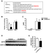
Figure 7. MOR1D C-Terminus is Critical for MIS and MOR1D/GRPR Dimerization
(A) Sequence comparison of MOR1D and MOR1 reveals a unique motif in MOR1D C-terminus. Synthesized peptide Tat-MOR1DCT contains a Tat domain from human immunodeficiency virus-type 1 and the motif from MOR1DCT. Control peptide contains Tat domain and scrambled sequence of MOR1DCT. (B) Tat-MOR1DCT blocked MIS without affecting GIS. (C) Tat-MOR1DCT had no effect on MIA. (D and E) Co-IP by anti-MOR1D (D) and quantified O.D. ratio of GRPR and MOR1D (E) showing Tat-MOR1DCT decreased GRPR/MOR1D interaction in the lumbar spinal cord. In all experiments, n = 6~8 per group. *p < 0.05. Error bars represent standard error of the mean.

Comment in

  • Why does morphine make you itch?
    Miyamoto T, Patapoutian A. Miyamoto T, et al. Cell. 2011 Oct 14;147(2):261-2. doi: 10.1016/j.cell.2011.09.026. Cell. 2011. PMID: 22000005

Similar articles

Cited by

References

    1. Abbadie C, Pan Y, Drake CT, Pasternak GW. Comparative immunohistochemical distributions of carboxy terminus epitopes from the mu-opioid receptor splice variants MOR-1D, MOR-1 and MOR-1C in the mouse and rat CNS. Neuroscience. 2000;100:141–153. - PubMed
    1. Agnati LF, Ferre S, Lluis C, Franco R, Fuxe K. Molecular mechanisms and therapeutical implications of intramembrane receptor/receptor interactions among heptahelical receptors with examples from the striatopallidal GABA neurons. Pharmacol Rev. 2003;55:509–550. - PubMed
    1. Alvarez VA, Arttamangkul S, Dang V, Salem A, Whistler JL, Von Zastrow M, Grandy DK, Williams JT. mu-Opioid receptors: Ligand-dependent activation of potassium conductance, desensitization, and internalization. J Neurosci. 2002;22:5769–5776. - PMC - PubMed
    1. Andoh T, Yageta Y, Konno M, Yamaguchi-Miyamoto T, Takahata H, Nojima H, Nemoto H, Kuraishi Y. Evidence for separate involvement of different mu-opioid receptor subtypes in itch and analgesia induced by supraspinal action of opioids. J Pharmacol Sci. 2008;106:667–670. - PubMed
    1. Ballantyne JC, Loach AB, Carr DB. Itching after epidural and spinal opiates. Pain. 1988;33:149–160. - PubMed

Publication types

  NODES
admin 2
Association 1
INTERN 12
twitter 2