. 2011 Oct 28;147(3):525-38.
doi: 10.1016/j.cell.2011.10.001.

Distal airway stem cells yield alveoli in vitro and during lung regeneration following H1N1 influenza infection

Affiliations

Distal airway stem cells yield alveoli in vitro and during lung regeneration following H1N1 influenza infection

Pooja A Kumar et al. Cell. .

Abstract

The extent of lung regeneration following catastrophic damage and the potential role of adult stem cells in such a process remains obscure. Sublethal infection of mice with an H1N1 influenza virus related to that of the 1918 pandemic triggers massive airway damage followed by apparent regeneration. We show here that p63-expressing stem cells in the bronchiolar epithelium undergo rapid proliferation after infection and radiate to interbronchiolar regions of alveolar ablation. Once there, these cells assemble into discrete, Krt5+ pods and initiate expression of markers typical of alveoli. Gene expression profiles of these pods suggest that they are intermediates in the reconstitution of the alveolar-capillary network eradicated by viral infection. The dynamics of this p63-expressing stem cell in lung regeneration mirrors our parallel finding that defined pedigrees of human distal airway stem cells assemble alveoli-like structures in vitro and suggests new therapeutic avenues to acute and chronic airway disease.


Figures

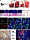
Fig. 1
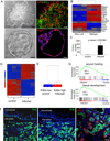
Fig. 1. Appearance of p63/Krt5-expressing cells in infected lung
A. Infected and control lung at 11dpi. Pulmonary edema and hemorrhage are evident in the infected lung. B. Viral M2 protein expression in lung tissue at progressive dpi (top panel). Lung histology at corresponding dpi (bottom panel). Graphics depicts overall trend of viral load versus lung tissue damage. Scale bar, 200µm. C. Detection of p63-expressing cells in sections of lung at 0dpi (top) and at 11dpi. Scale bar, 20µm. D. Co-localization of p63 and Krt5 expression in “pods” in lung parenchyma at 15dpi. Br, bronchiole. Scale bar, 20µm. E. Localization of Krt5 (brown) at 15dpi. Br, bronchiole. Scale bar, 100µm. F. Detection of BrdU (green) and Krt5 (red) at 15dpi after 4 days of BrdU labelling. Br, bronchiole. Scale bar, 100µm.
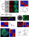
Fig. 2
Fig. 2. Cloning and pedigree tracking of regiospecific stem cells from human lung
A. Schematic of human airways as source of cells for stem cell cloning. NESCs, nasal epithelial cells; TASCs, tracheobronchial epithelial cells; DASCs, small airway epithelial cells. Left panel, Epithelial cell clones on irradiated Swiss 3T3 cells. Middle panel, p63 immunofluorescence. Right panel, Keratin 5 (Krt5) immunofluorescence. Scale bar, 50µm. B. Comparative heatmap of NESC, TASC and DASC expression profiles. C. Principle Component Analysis (PCA) of expression microarrays. D. Schematic showing pedigree tracking and expansion. E. Air-liquid interface (ALI) differentiation of NESCs indicated by antibodies to tubulin and mucin 5A to mark ciliated cells and goblet cells, respectively. Histogram of counts of ciliated cells (tub) and goblet cells (muc). Expression heatmap comparing NESCs grown on 3T3 feeder cells (NESC-3T3) with those differentiated in ALI culture. Scale bar, 20µm. F. Self-assembly sphere (SAS) assay of NESCs. Staining SASs with antibodies to tubulin (green) and Muc5A (red) revealing ciliated cells and goblet cells, respectively. Heatmap depicting differential gene expression of NESCs and those differentiated in SAS culture. Scale bar, 50µm. G. 21 day 3-D Matrigel cultures of NESCc (phase contrast, upper left panel) and confocal immunofluorescence using antibodies to involucrin and Krt10 (upper right panel and lower left panel). Scale bar, 50µm. Gene expression heatmap of NESC and those in Matrigel (lower right panel). H. PCA of whole genome expression microarrays of NESCs and those differentiated by indicated methods.
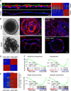
Fig. 3
Fig. 3. Alveolar-like structures from DASCs in 3-D Matrigel cultures
A. TASCs and DASCs following 21 days of ALI culture using antibodies to tubulin (green) and Muc5A (red) to reveal ciliated cells and goblet cells, respectively. Scale bar, 20µm. Heatmap of gene expression differences in DASC and TASC lines upon differentiation in ALI cultures. B. Comparison of structures formed by TASCs and DASCs following 21 days in 3-D Matrigel. Top panel, Phase contrast and confocal images of TASC spheres. Confocal immunoflourescence with antibodies to the squamous marker Krt10 (red). Scale bar, 20µm. Bottom panel, Multivesicular, unilaminar structures formed by DASCs following 21 days in Matrigel and confocal immunofluorescence image showing PDPN immunofluorescence. Scale bar, 50µm. C. Top panel, Immunofluorescence micrographs of staining patterns on human deep lung with anti-alveoli monoclonal antibody 4C10. Scale bar, 100µm. Bottom panel, Immunofluorescence micrographs of monoclonal antibody 4C10 on the structures developed from DASC lines in 21-day Matrigel cultures. Scale bar, 50µm. D. Heatmap of differentially expressed genes from microarrays of DASC and TASC lines differentiated in 3-D Matrigel. E. Heatmap of DASC genes that are both differentially expressed in Matrigel and expressed in interstitial regions of mouse lung. F. Gene Set Enrichment Analysis of datasets derived from TASC and DASC differentiation in 3-D Matrigel culture.
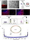
Fig. 4
Fig. 4. Conservation of alveoli-like differentiation in rat DASC pedigrees
A. Rat DASCs on 3T3 cells derived from single cell suspension of deep lung tissue. Top left, Phase contrast; Bottom left, Immunofluorescence with anti-p63 antibodies (red). Middle panel, Image of unilaminar structures produced by rat DASC pedigree-specific lines after 21 days in 3-D Matrigel culture. Scale bar, 50µm. B. Schematic showing immunization of mice with rat lung and screening for interstitial lung-specific monoclonal antibodies. C. Immunofluorescence on rat deep lung and unilaminar structures developed from rat DASC lines in Matrigel cultures with anti-alveoli monoclonal antibodies 13A1 and 54D1. Scale bar, 100µm. D. Schematic of the differentiation potential of regiospecific airway stem cells derived from multiple in vitro models. E. Venn diagram depicting differentially expressed genes between TASC and DASC stem cell pedigrees, while the graphics below indicate the absolute fold-change in gene expression between TASC and DASC among more than 17,000 informative genes.
Fig. 5
Fig. 5. Expansion of basal-like cell population in lung following infection
A. Upper left, Image of typical colony of basal-like cells derived from three-week-old mouse lung. Scale bar, 200µm. Upper right, Immunofluorescence image of typical colony stained with anti-Krt5 (green) and anti-p63 (red). Scale bar, 50µm. Lower left, Image of spheres generated by growth of cloned cells in 3-D Matrigel culture for 10 days showing hollow lumen. Scale bar, 50µm. Lower right, Section of 3-D sphere stained with anti-Aqp5 antibodies. Scale bar, 50µm. B. Heatmap showing relative gene expression pattern of selected genes in cloned stem cells and 3-D Matrigel spheres. C. Quantification of colony numbers obtained from control and infected mice at 12 dpi. Number of mice n=7. Error bars, SD of mean. D. Heatmap of differentially expressed genes (> two-fold) between three different colonies obtained from control and infected lungs. E. Unsupervised clustering analysis using whole transcriptome for control and infected colonies. F. Immunofluorescence micrographs showing distribution of Krt5 (left panel; green), Krt6 (middle panel; green), and p63 (red) and Krt6 (green) in sections of 12dpi lung (right panel). Bronchiolar and interstitial lung regions are marked. Scale bar, 20µm. G.GSEA survey data showing enrichment of wound healing gene expression in colonies derived from infected lung.
Fig. 6
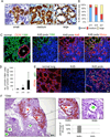
Fig. 6. Dynamics of Krt5 pods in infected lung parenchyma
A. Sections of infected lung showing variable appearance of Krt5 pods revealed by Krt5 immunohistochemistry. Scale bar, 20µm. B. Quantification of the Krt5 pod size categories at 11, 15, and 21dpi. Number of clusters (n) counted at 11, 15 and 21 dpi are 200, 424 and 572, respectively. C. Left panel, Staining of normal lung with the 11B6 monoclonal antibody showing alveolar-specific signal. Middle panels, Staining of Krt5 pods in damaged lung with the 11B6 monoclonal antibody (green) and Krt5 antibodies (red), respectively. Right panel, A merge of 11B6 and Krt5 antibody signals of the middle panels. D. Quantification of Krt5+11B6+ cells per Krt5+ cell area over indicated dpi. Number of mice n=4. Error bars indicate standard deviation of the mean. Scale bar, 50µm. E. Staining of normal lung with antibodies to PDPN (red) (left panel). Middle panel, Staining of region of damaged lung lacking Krt5 pods with antibodies to PDPN (red) and p63 (green) showing an absence of signal. Right panel, Co-localization of PDPN (red) and p63 (green) in region of damaged lung that has clusters of Krt5 pods. Scale bar, 50µm. F. Section of 12dpi lung showing clusters of Krt5 pods about bronchioles that possess Krt5-expressing cells (red star) and the absence of Krt5 pods surrounding bronchioles lacking Krt5-expressing cells (green star). Scale bar, 100µm. Histogram reports direct counts of peribronchiolar clusters of Krt5 pods adjacent to bronchioles with (93.8%) and without Krt5-expressing cells. Number of sections n=32. Error bars, SD of mean.
Fig. 7
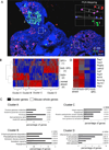
Fig. 7. Gene expression profiles of regionally distinct infected lung
A. Section of 25dpi lung stained with antibodies to Krt5 (green), SPC (red), and counterstained with DAPI (blue). Four regions are demarcated with boxes as laser capture microdissection _targets 1) SPC+ cells in densely infiltrated zones, 2) Krt5 pods, 3) SPC−/Krt5− zones with dense infiltrates, and 4) SPC+ cells in normal lung. Inset shows PCA of three independent LCM samples corresponding to regions 1–4. Scale bar, 200µm. B. Heatmap of 2205 differentially expressed genes with p value < 0.05 derived from LCM samples of regions 1–4. Gene clusters A–D are indicated. C. Gene Ontology analysis of gene clusters A–D indicated in heatmap together with associated p values. D. Heatmap indicating relative expression of individual genes linked to alveoli in the datasets corresponding to regions 1–4.
Fig. 8
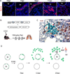
Fig. 8. Lineage tracing and schematic of response of Krt5 cells to influenza infection
A. Localization of Krt14 expression in influenza-infected lung at indicated dpi. Br, bronchiole. Scale bar, 20µm. B. Schematic of alleles for conditional expression of β-galactosidase via Krt14-Cre recombinase activation. C. LacZ staining (blue) and Krt5 immunochemistry (brown) in 25dpi mouse lung. Scale bar, 20µm. D. Schematic depicting two bronchioles (Br) with rare Krt5-expressing cells at the basement membrane that proliferate in response to infection (top) or are killed by the infection (bottom). Early Krt5 pods are evident by 11dpi, and mature with luminal formation by day 25. Major questions for how they link to alveolar capillary (av) beds and to the bronchioles are indicated.

Comment in

Similar articles

Cited by

References

    1. Barrandon Y, Green H. Three clonal types of keratinocyte with different capacities for multiplication. Proc. Natl. Acad. Sci. U.S.A. 1987;84:2302–2306. - PMC - PubMed
    1. Belser JA, Szretter KJ, Katz JM, Tumpey TM. Use of animal models to understand the pandemic potential of highly pathogenic avian influenza viruses. Adv. Virus Res. 2009;73:55–97. - PubMed
    1. Berthiaume Y, Lesur O, Dagenais A. Treatment of adult respiratory distress syndrome: plea for rescue therapy of the alveolar epithelium. Thorax. 1999;54:150–160. - PMC - PubMed
    1. Cole BB, Smith RW, Jenkins KM, Graham BB, Reynolds PR, Reynolds SD. Tracheal basal cells: a facultative progenitor cell pool. Am. J. Pathol. 2010;177:362–376. - PMC - PubMed
    1. Eaton DC, Helms MN, Koval M, Bao HF, Jain L. The contribution of epithelial sodium channels to alveolar function in health and disease. Annu Rev Physiol. 2009;71:403–423. - PubMed

Publication types

Substances

Associated data

  NODES
Project 1
twitter 2