Identification of the novel localization of tenascinX in the monkey choroid plexus and comparison with the mouse
- PMID: 22073359
- PMCID: PMC3167336
- DOI: 10.4081/ejh.2009.e27
Identification of the novel localization of tenascinX in the monkey choroid plexus and comparison with the mouse
Abstract
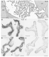
Tenascin-X (Tn-X) belongs to the tenascin family of glycoproteins and has been reported to be significantly associated with schizophrenia in a single nucleotide polymorphism analysis in humans. This finding indicates an important role of Tn-X in the central nervous system (CNS). However, details of Tn-X localization are not clear in the primate CNS. Using immunohistochemical techniques, we found novel localizations of Tn-X in the interstitial connective tissue and around blood vessels in the choroid plexus (CP) in macaque monkeys. To verify the reliability of Tn-X localization, we compared the Tn-X localization with the tenascin-C (Tn-C) localization in corresponding regions using neighbouring sections. Localization of Tn-C was not observed in CP. This result indicated consistently restricted localization of Tn-X in CP. Comparative investigations using mouse tissues showed equivalent results. Our observations provide possible insight into specific roles of Tn-X in CP for mammalian CNS function.
Keywords: Ehlers-Danlos syndrome; choroid plexus; monkey; mouse; schizophrenia.; tenascin-X.
Figures


Similar articles
-
Novel localization of tenascin-X in adult mouse leptomeninges and choroid plexus.Ann Anat. 2008;190(4):324-8. doi: 10.1016/j.aanat.2008.04.003. Epub 2008 May 27. Ann Anat. 2008. PMID: 18595676
-
Tenascin-X: beyond the architectural function.Cell Adh Migr. 2015;9(1-2):154-65. doi: 10.4161/19336918.2014.994893. Cell Adh Migr. 2015. PMID: 25793578 Free PMC article. Review.
-
Differential expression of tenascin-C and tenascin-X in human astrocytomas.Acta Neuropathol. 1997 May;93(5):431-7. doi: 10.1007/s004010050636. Acta Neuropathol. 1997. PMID: 9144580
-
Involvement of chondroitin sulfates on brain-derived tenascin-R in carbohydrate-dependent interactions with fibronectin and tenascin-C.Brain Res. 2000 Apr 28;863(1-2):42-51. doi: 10.1016/s0006-8993(00)02075-8. Brain Res. 2000. PMID: 10773191
-
The tenascin family of ECM glycoproteins: structure, function, and regulation during embryonic development and tissue remodeling.Dev Dyn. 2000 Jun;218(2):235-59. doi: 10.1002/(SICI)1097-0177(200006)218:2<235::AID-DVDY2>3.0.CO;2-G. Dev Dyn. 2000. PMID: 10842355 Review.
Cited by
-
Effects of Ethanol on Brain Extracellular Matrix: Implications for Alcohol Use Disorder.Alcohol Clin Exp Res. 2016 Oct;40(10):2030-2042. doi: 10.1111/acer.13200. Epub 2016 Sep 1. Alcohol Clin Exp Res. 2016. PMID: 27581478 Free PMC article. Review.
-
Histochemistry through the years, browsing a long-established journal: novelties in traditional subjects.Eur J Histochem. 2010 Dec 16;54(4):e51. doi: 10.4081/ejh.2010.e51. Eur J Histochem. 2010. PMID: 21263750 Free PMC article.
-
Tenascin-X-Discovery and Early Research.Front Immunol. 2021 Jan 11;11:612497. doi: 10.3389/fimmu.2020.612497. eCollection 2020. Front Immunol. 2021. PMID: 33505400 Free PMC article. Review. No abstract available.
-
Extracellular matrix functions during neuronal migration and lamination in the mammalian central nervous system.Dev Neurobiol. 2011 Nov;71(11):889-900. doi: 10.1002/dneu.20946. Dev Neurobiol. 2011. PMID: 21739613 Free PMC article. Review.
-
Tenascin-X, Congenital Adrenal Hyperplasia, and the CAH-X Syndrome.Horm Res Paediatr. 2018;89(5):352-361. doi: 10.1159/000481911. Epub 2018 May 7. Horm Res Paediatr. 2018. PMID: 29734195 Free PMC article. Review.
References
-
- Aase K, Lymboussaki A, Kaipainen A, Olofsson B, Alitalo K, Eriksson U. Localization of VEGF-B in the mouse embryo suggests a paracrine role of the growth factor in the developing vasculature. Dev Dyn. 1999;215:1–25. - PubMed
-
- Adams JC, Watt FM. Regulation of development and differentiation by the extracellular matrix. Development. 1993;117:1183–98. - PubMed
-
- Ballard VLT, Sharma A, Duignan I, Holm JM, Chin A, Choi R, Hajjar KA, Wong SC, Edelberge JM. Vascular tenascin-C regulates cardiac endothelial phenotype and neovascularization. FASEB J. 2006;20:717–9. - PubMed
-
- Borlongan CV, Skinner SJM, Geaney M, Vasconcellos AV, Elliott RB, Emerich DF. Intracerebral transplantation of porcine choroid plexus provides structural and functional neuroprotection in a rodent model of stroke. Stroke. 2004;35:2206–10. - PubMed
Publication types
MeSH terms
Substances
LinkOut - more resources
Full Text Sources
Miscellaneous

