. 2012 Jan 4;31(1):14-28.
doi: 10.1038/emboj.2011.423. Epub 2011 Nov 25.

IAPs regulate the plasticity of cell migration by directly _targeting Rac1 for degradation

Affiliations

IAPs regulate the plasticity of cell migration by directly _targeting Rac1 for degradation

Tripat Kaur Oberoi et al. EMBO J. .

Abstract

Inhibitors of apoptosis proteins (IAPs) are a highly conserved class of multifunctional proteins. Rac1 is a well-studied Rho GTPase that controls numerous basic cellular processes. While the regulation of nucleotide binding to Rac1 is well understood, the molecular mechanisms controlling Rac1 degradation are not known. Here, we demonstrate X-linked IAP (XIAP) and cellular IAP1 (c-IAP1) directly bind to Rac1 in a nucleotide-independent manner to promote its polyubiquitination at Lys147 and proteasomal degradation. These IAPs are also required for degradation of Rac1 upon CNF1 toxin treatment or RhoGDI depletion. Consistently, downregulation of XIAP or c-IAP1 by various strategies led to an increase in Rac1 protein levels in primary and tumour cells, leading to an elongated morphology and enhanced cell migration. Further, XIAP counteracts Rac1-dependent cellular polarization in the developing zebrafish hindbrain and promotes the delamination of neurons from the normal tissue architecture. These observations unveil an evolutionarily conserved role of IAPs in controlling Rac1 stability thereby regulating the plasticity of cell migration and morphogenesis.


Conflict of interest statement

The authors declare that they have no conflict of interest.

Figures
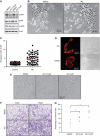
Figure 1
Figure 1
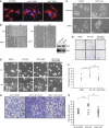
Depletion of IAPs with IAC induces elongated morphology and migration. (A) HeLa cells were treated with two concentrations of IAC for 16 h and the depletion of various IAPs was monitored by immunoblots. (B) Phase-contrast images of HeLa cells grown on plastic dishes coated with gelatin. The cells were seeded in the presence of DMSO or IAC (5 μM) for 15 h. (C) Elongation index of cells shown in (B). (D) HeLa cells were transfected with LifeAct to stain polymerized actin in live cells. The cells were seeded onto collagen-coated plates and the morphological changes were observed by time-lapse microscopy. (E) Morphological changes of HeLa cells embedded in thick collagen matrices 24 h after IAC treatment. (F) HeLa cells were allowed to migrate in a transwell migration chamber after IAC treatment. The migrated cells were then fixed and stained with crystal violet. Data from one representative experiment are shown. (G) The quantifications of transwell migration experiments from three independent experiments are shown (*P<0.05).
Figure 2
Figure 2
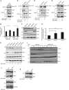
IAPs regulate the stability of Rac1. (A) HeLa cells were treated either with IAC (2.5 μM) or with DMSO for 16 h and the active GTP-bound form of active Rac1 or RhoA (C) was precipitated as mentioned in Materials and methods. (B) Quantification of total and active Rac1 levels (by densitometry as described in Materials and methods) in control and IAC-treated cells is shown. Data from three independent experiments are shown. Error bars represent ±s.d. of the mean. The depletion of c-IAP1 and the total levels of Rac1 and RhoA were monitored in the total lysates. (D) HeLa cells were transfected with c-IAP1 or (E) XIAP siRNAs for 48 h and the amount of GTP-bound Rac1 was precipitated. The levels of total Rac1 were monitored in the cell lysates by immunoblots. (F) HeLa cells were transfected with various siRNAs for 48 h. Total cell lysates were western blotted for monitoring the protein levels of various IAPs and Rac1. (G) The increase in Rac1 levels was quantified by densitometry. The data from three independent experiments are shown (*P<0.05, **P<0.01, ***P<0.005). Error bars represent ±s.d. of the mean. (H) 293T cells were transfected with two sets of XIAP and three sets of c-IAP1 siRNAs. Rac1 levels were monitored by western blots. Quantification of Rac1 increase is provided. (I) Combined loss of XIAP and c-IAP1 enhances the protein half-life of Rac1. HeLa cells transfected with XIAP siRNAs were treated with IAC and cycloheximide was added to cells to monitor the protein half-life of Rac1. (J) Western blot analyses showing protein levels of Rac1 and Ras in wild-type (WT) and c-IAP1−/− or (K) WT and XIAP−/− MEFs. The signalling intensities of Rac1 and Ras bands were quantified by densitometry and normalized to actin levels. Figure source data can be found in Supplementary data.
Figure 3
Figure 3
Rac1 is required for IAC-mediated elongated morphology and cell migration. (A) Representative images from wild-type, c-IAP1−/− and XIAP−/− MEFs grown on glass and stained with Phalloidin-TRITC (red). (B) Phase-contrast images at 0 and 8 or 10 h from a representative wound healing experiment performed with wild-type and XIAP−/− or c-IAP1−/− MEFs. (C) Stable cell lines expressing Rac1 or control shRNAs were treated with IAC and the morphological changes were monitored by phase-contrast microscopy. The representative fields from each condition as well as western blot showing the knockdown efficiency of Rac1 shRNA are shown. (D) HeLa cells seeded onto gelatin were treated with DMSO, IAC or IAC+NSC-23766 (50 μM) and observed under a microscope at various time intervals. Cropped snap shots from Supplementary movies are shown. (E) HeLa cells were pretreated with Rac1 inhibitor NSC-23766 and the influence of IAC in mediating transwell migration was monitored. (F) The transwell migration experiments shown in (E) were quantified by computer-assisted algorithms as mentioned in Materials and methods. The data from three independent experiments are shown (**P<0.01, ***P<0.005). (G) HeLa cells were pretreated with control or Rac1 siRNAs and the influence of IAC in mediating transwell migration was monitored. (H) The transwell migration experiments shown in (G) were quantified by computer-assisted algorithms as mentioned in Materials and methods. The data from three independent experiments are shown (**P<0.01, ***P<0.005).
Figure 4
Figure 4
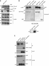
IAPs can bind to Rac1 in vitro and in vivo. (A) Endogenous interaction between Rac1 and IAPs. HeLa cells grown in 10 cm dishes were stimulated with HGF (100 ng, 15 h) and lysed with appropriate buffer. Endogenous Rac1 was then immunoprecipitated and the immunocomplexes were resolved in an SDS–PAGE and western blotted for co-precipitating XIAP and c-IAP1. (B) In-vitro binding assays with purified proteins showing XIAP and c-IAP1 binding to GST–Rac1 in a GTP/GDP-independent manner. The GST–Rac1 is pretreated with GTPγS and GDP to analyse the dependency of nucleotides in binding with IAPs. The nucleotide loading was confirmed by PAK1-PBD pull-down experiments, which revealed the GTP-specific binding. (C) The interaction between Myc-tagged XIAP or XIAP (1–240) and HA–Rac1 is tested after expressing them in 293T cells. The unspecific binding of antibodies in immunoprecipitation experiments was always checked with respective isotype controls. Figure source data can be found in Supplementary data.
Figure 5
Figure 5
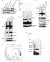
Binding of IAPs to Rac1 promotes Rac1 polyubiquitination and degradation. (A) The rescue in the levels of Rac1 under the influence of proteasomal inhibitors MG132 upon IAP overexpression is monitored by western blotting. (B) c-IAP1 directly ubiquitinates Rac1. Purified recombinant Rac1 proteins with various mutations were subjected to in-vitro ubiquitination with c-IAP1 as described in Materials and methods. The conjugation of ubiquitin to Rac1 was monitored by immunoblots. The lower exposure of Rac1 was shown below. (C) XIAP and c-IAP1 promoted the polyubiquitination of activated Rac1. 293T cells were transfected with Myc–Rac1Q61L or Myc–Rac1Q61LK147R in combination with Flag–IAPs and HA–ubiquitin constructs. Rac1Q61L was immunoprecipitated by using Myc antibody and the immunocomplexes were western blotted for HA (ubiquitin), Rac1 and Flag for IAPs. The cells were treated with MG132 for 6 h before lysing. (D) Ubiquitination of Myc–Rac1Q61L by c-IAP1 and c-IAPH588A in 293T cells is analysed as mentioned in (C). (E) c-IAP1 directly ubiquitinates Rac1 at lysine 147. Recombinant Rac1Q61L was subjected to an in-vitro ubiquitination reaction as mentioned in Materials and methods with c-IAP1. The modification of Rac1 protein was monitored by immunoblots. (F) Samples from in-vitro ubiquitination reaction of Rac1 with c-IAP1 (E) were subjected to mass spectrometric analysis after digestion with trypsin. MS/MS spectrum of the Rac1 peptide carrying Gly-Gly modification at position K147 is shown. The mass of the triply charged precursor ion at 744.41 was measured with a mass deviation of 0.08 p.p.m. Arrow indicates the gel band where the peptide was detected. Figure source data can be found in Supplementary data.
Figure 6
Figure 6
IAPs are required for polyubiquitination and proteasomal degradation of endogenous Rac1. (A) HeLa cells were transfected with control, XIAP and RhoGDI siRNAs. The levels of various proteins were monitored by immunoblots. (B) HeLa cells were transfected with control, RhoGDI or RhoGDI and XIAP siRNAs. Endogenous Rac1 was immunoprecipitated and the conjugation of polyubiquitin chains to Rac1 was monitored by immunoblots. The cells were treated with MG132 for 6 h before lysing to preserve ubiquitinated Rac1. (C) IAPs are required for CNF1-mediated Rac1 degradation. 293T cells transfected with siRNAs and/or treated with IAC are further treated with 200 ng/ml of CNF1 toxin for 6 h. The levels of Rac1 and IAPs are monitored. (D) 293T cells transfected with control or XIAP siRNAs+IAC are treated with CNF1 toxin as mentioned earlier. Endogenous Rac1 was immunoprecipitated and conjugation of ubiquitin is monitored. The cells were treated with MG132 for 6 h before lysing. Figure source data can be found in Supplementary data.
Figure 7
Figure 7
Expression of dnRac1 or XIAP in the hindbrain rhombic lip causes cells to delaminate into the ventricle. (A) Lateral view of the cerebellum and anterior hindbrain of 48 h.p.f. Tg(atoh1a:Gal4TA4)hzm2 embryos expressing FynTagRFP-T, FynTagRFP-T/Rac1, FynTagRFP-T/dnRac1 or FynTagRFP-T/XIAP. The cells were found either as isolated cells (arrowheads) or as clusters (arrows) in the cerebrospinal fluid of the ventricle. Abbreviations: 4th V; 4th ventricle; A, anterior; cb, cerebellum; D, dorsal; hb, hindbrain; P, posterior; V, ventral. (B) Graph showing the percentage of embryos in each condition with one or more abnormal TagRFP-T-expressing cell cluster. (C) Graph showing the percentage of embryos with one or more isolated, rounded TagRFP-T-expressing cells. For both graphs, n=6 independent experiments, one-way ANOVA and Tukey’s post hoc test. **P<0.01, *P<0.05. Error bars represent ±s.d. (D) Zebrafish XIAP binds to Rac1. Dr-XIAP produced in rabbit reticulolysates was subjected to GST pull-down experiments with either GST or GST–RacQ61L or with GST–RacT17N. (E) Dr-XIAP and Rac1 constructs were expressed in 293T cells and the degradation of Rac1 was monitored in the immunoblots. (F) Rac1 was immunoprecipitated from immortalized cerebellar granule neurons from mice and the co-precipitation of XIAP was monitored by immunoblots. (G) mCGN cells were transfected with XIAP siRNA and the increase in total Rac1 levels was monitored by immunoblots. (H) Proposed model for IAP-mediated degradation of Rac1. GEFs and GAPs control the binding of GTP/GDP to Rac1. XIAP–c-IAP1 complex directly binds to Rac1 and promote the conjugation of polyubiquitin chains to Lysine147 of activated Rac1 in vivo. Loss of IAPs stabilizes Rac1 protein leading to elongated/mesenchymal mode of migration in normal and tumour cells. Figure source data can be found in Supplementary data.

Comment in

Similar articles

Cited by

References

    1. Bertrand MJ, Doiron K, Labbe K, Korneluk RG, Barker PA, Saleh M (2009) Cellular inhibitors of apoptosis cIAP1 and cIAP2 are required for innate immunity signaling by the pattern recognition receptors NOD1 and NOD2. Immunity 30: 789–801 - PubMed
    1. Blankenship JW, Varfolomeev E, Goncharov T, Fedorova AV, Kirkpatrick DS, Izrael-Tomasevic A, Phu L, Arnott D, Aghajan M, Zobel K, Bazan JF, Fairbrother WJ, Deshayes K, Vucic D (2008) Ubiquitin binding modulates IAP antagonist stimulated proteasomal degradation of c IAP1 and c IAP2. Biochem J 417: 1–3 - PubMed
    1. Boulter E, Garcia-Mata R, Guilluy C, Dubash A, Rossi G, Brennwald PJ, Burridge K (2010) Regulation of Rho GTPase crosstalk, degradation and activity by RhoGDI1. Nat Cell Biol 12: 477–483 - PMC - PubMed
    1. Cartier J, Berthelet J, Marivin A, Gemble S, Edmond V, Plenchette S, Lagrange B, Hammann A, Dupoux A, Delva L, Eymin B, Solary E, Dubrez L (2011) Cellular inhibitor of apoptosis protein-1 (CIAP1) can regulate E2F1-mediated control of cyclin transcription. J Biol Chem 29: 26406–26417 - PMC - PubMed
    1. Castillo-Lluva S, Tatham MH, Jones RC, Jaffray EG, Edmondson RD, Hay RT, Malliri A (2010) SUMOylation of the GTPase Rac1 is required for optimal cell migration. Nat Cell Biol 12: 1078–1085 - PMC - PubMed

Publication types

Substances

  NODES
twitter 2