Boswellia sacra essential oil induces tumor cell-specific apoptosis and suppresses tumor aggressiveness in cultured human breast cancer cells
- PMID: 22171782
- PMCID: PMC3258268
- DOI: 10.1186/1472-6882-11-129
Boswellia sacra essential oil induces tumor cell-specific apoptosis and suppresses tumor aggressiveness in cultured human breast cancer cells
Abstract
Background: Gum resins obtained from trees of the Burseraceae family (Boswellia sp.) are important ingredients in incense and perfumes. Extracts prepared from Boswellia sp. gum resins have been shown to possess anti-inflammatory and anti-neoplastic effects. Essential oil prepared by distillation of the gum resin traditionally used for aromatic therapy has also been shown to have tumor cell-specific anti-proliferative and pro-apoptotic activities. The objective of this study was to optimize conditions for preparing Boswellea sacra essential oil with the highest biological activity in inducing tumor cell-specific cytotoxicity and suppressing aggressive tumor phenotypes in human breast cancer cells.
Methods: Boswellia sacra essential oil was prepared from Omani Hougari grade resins through hydrodistillation at 78 or 100 °C for 12 hours. Chemical compositions were identified by gas chromatography-mass spectrometry; and total boswellic acids contents were quantified by high-performance liquid chromatography. Boswellia sacra essential oil-mediated cell viability and death were studied in established human breast cancer cell lines (T47D, MCF7, MDA-MB-231) and an immortalized normal human breast cell line (MCF10-2A). Apoptosis was assayed by genomic DNA fragmentation. Anti-invasive and anti-multicellular tumor properties were evaluated by cellular network and spheroid formation models, respectively. Western blot analysis was performed to study Boswellia sacra essential oil-regulated proteins involved in apoptosis, signaling pathways, and cell cycle regulation.
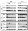
Results: More abundant high molecular weight compounds, including boswellic acids, were present in Boswellia sacra essential oil prepared at 100 °C hydrodistillation. All three human breast cancer cell lines were sensitive to essential oil treatment with reduced cell viability and elevated cell death, whereas the immortalized normal human breast cell line was more resistant to essential oil treatment. Boswellia sacra essential oil hydrodistilled at 100 °C was more potent than the essential oil prepared at 78 °C in inducing cancer cell death, preventing the cellular network formation (MDA-MB-231) cells on Matrigel, causing the breakdown of multicellular tumor spheroids (T47D cells), and regulating molecules involved in apoptosis, signal transduction, and cell cycle progression.
Conclusions: Similar to our previous observations in human bladder cancer cells, Boswellia sacra essential oil induces breast cancer cell-specific cytotoxicity. Suppression of cellular network formation and disruption of spheroid development of breast cancer cells by Boswellia sacra essential oil suggest that the essential oil may be effective for advanced breast cancer. Consistently, the essential oil represses signaling pathways and cell cycle regulators that have been proposed as therapeutic _targets for breast cancer. Future pre-clinical and clinical studies are urgently needed to evaluate the safety and efficacy of Boswellia sacra essential oil as a therapeutic agent for treating breast cancer.
Figures


Similar articles
-
Frankincense essential oil prepared from hydrodistillation of Boswellia sacra gum resins induces human pancreatic cancer cell death in cultures and in a xenograft murine model.BMC Complement Altern Med. 2012 Dec 13;12:253. doi: 10.1186/1472-6882-12-253. BMC Complement Altern Med. 2012. PMID: 23237355 Free PMC article.
-
Frankincense oil derived from Boswellia carteri induces tumor cell specific cytotoxicity.BMC Complement Altern Med. 2009 Mar 18;9:6. doi: 10.1186/1472-6882-9-6. BMC Complement Altern Med. 2009. PMID: 19296830 Free PMC article.
-
Comparative Analysis of Pentacyclic Triterpenic Acid Compositions in Oleogum Resins of Different Boswellia Species and Their In Vitro Cytotoxicity against Treatment-Resistant Human Breast Cancer Cells.Molecules. 2019 Jun 7;24(11):2153. doi: 10.3390/molecules24112153. Molecules. 2019. PMID: 31181656 Free PMC article.
-
Frankincense--therapeutic properties.Postepy Hig Med Dosw (Online). 2016 Jan 4;70:380-91. doi: 10.5604/17322693.1200553. Postepy Hig Med Dosw (Online). 2016. PMID: 27117114 Review.
-
Pharmacological evidences for cytotoxic and antitumor properties of Boswellic acids from Boswellia serrata.J Ethnopharmacol. 2016 Sep 15;191:315-323. doi: 10.1016/j.jep.2016.06.053. Epub 2016 Jun 21. J Ethnopharmacol. 2016. PMID: 27346540 Review.
Cited by
-
Chemistry and biology of essential oils of genus boswellia.Evid Based Complement Alternat Med. 2013;2013:140509. doi: 10.1155/2013/140509. Epub 2013 Mar 6. Evid Based Complement Alternat Med. 2013. Retraction in: Evid Based Complement Alternat Med. 2014;2014:605304. doi: 10.1155/2014/605304 PMID: 23533463 Free PMC article. Retracted.
-
Frankincense essential oil prepared from hydrodistillation of Boswellia sacra gum resins induces human pancreatic cancer cell death in cultures and in a xenograft murine model.BMC Complement Altern Med. 2012 Dec 13;12:253. doi: 10.1186/1472-6882-12-253. BMC Complement Altern Med. 2012. PMID: 23237355 Free PMC article.
-
Exploring the Potent Anticancer Activity of Essential Oils and Their Bioactive Compounds: Mechanisms and Prospects for Future Cancer Therapy.Pharmaceuticals (Basel). 2023 Jul 31;16(8):1086. doi: 10.3390/ph16081086. Pharmaceuticals (Basel). 2023. PMID: 37631000 Free PMC article. Review.
-
Differential effects of selective frankincense (Ru Xiang) essential oil versus non-selective sandalwood (Tan Xiang) essential oil on cultured bladder cancer cells: a microarray and bioinformatics study.Chin Med. 2014 Jul 2;9:18. doi: 10.1186/1749-8546-9-18. eCollection 2014. Chin Med. 2014. PMID: 25006348 Free PMC article.
-
Innovative microwave-assisted biosynthesis of copper oxide nanoparticles loaded with platinum(ii) based complex for halting colon cancer: cellular, molecular, and computational investigations.RSC Adv. 2024 Jan 29;14(6):4005-4024. doi: 10.1039/d3ra08779d. eCollection 2024 Jan 23. RSC Adv. 2024. PMID: 38288146 Free PMC article.
References
-
- Maloney GA. Gold, frankincense, and myrrh : an introduction to Eastern Christian spirituality. New York: Crossroads Pub. Co; 1997.
MeSH terms
Substances
LinkOut - more resources
Full Text Sources
Other Literature Sources
Medical
Miscellaneous

