. 2012 Feb 14;22(2):320-33.
doi: 10.1016/j.devcel.2011.12.014. Epub 2012 Jan 25.

Role of PINK1 binding to the TOM complex and alternate intracellular membranes in recruitment and activation of the E3 ligase Parkin

Affiliations

Role of PINK1 binding to the TOM complex and alternate intracellular membranes in recruitment and activation of the E3 ligase Parkin

Michael Lazarou et al. Dev Cell. .

Abstract

Mutations in the mitochondrial kinase PINK1 and the cytosolic E3 ligase Parkin can cause Parkinson's disease. Damaged mitochondria accumulate PINK1 on the outer membrane where, dependent on kinase activity, it recruits and activates Parkin to induce mitophagy, potentially maintaining organelle fidelity. How PINK1 recruits Parkin is unknown. We show that endogenous PINK1 forms a 700 kDa complex with the translocase of the outer membrane (TOM) selectively on depolarized mitochondria whereas PINK1 ectopically _targeted to the outer membrane retains association with TOM on polarized mitochondria. Inducibly _targeting PINK1 to peroxisomes or lysosomes, which lack a TOM complex, recruits Parkin and activates ubiquitin ligase activity on the respective organelles. Once there, Parkin induces organelle selective autophagy of peroxisomes but not lysosomes. We propose that the association of PINK1 with the TOM complex allows rapid reimport of PINK1 to rescue repolarized mitochondria from mitophagy, and discount mitochondrial-specific factors for Parkin translocation and activation.


Figures

Figure 1
Figure 1
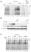
In vitro import and BN-PAGE analysis of PINK1. (A) [35S]-PINK1 was incubated with isolated HeLa mitochondria with or without 1 μM CCCP for increasing times as indicated. Samples were treated with or without Proteinase K (PK) and solubilized in 1% digitonin containing buffer. Mock import samples lacking mitochondria were treated as above as indicated. (B) Mitochondria were isolated from HeLa cells that were either untreated or treated with CCCP or vehicle control (DMSO). Isolated mitochondria were treated with or without external protease (PK) and subjected to BN-PAGE and immunoblotting using antibodies against PINK1 (outer membrane), Tom22 (outer membrane) and Complex II (inner membrane). (C) Radiolabeled WT PINK1 and PINK1 patient mutants A168P, H271Q and G309D were imported into isolated HeLa mitochondria as in (A). Radiolabeled proteins were detected by phosphorimage analysis. See also Figure S1.
Figure 2
Figure 2
PINK1 and Parkin complex analysis. (A-B) WT HeLa or YFP-Parkin HeLa cells were treated as indicated and subjected to BN-PAGE (A) or SDS-PAGE (B) and immunoblotted using α-PINK1 and either α-Tom20 (A) or α-actin (B) antibodies. (C) YFP-Parkin HeLa cells treated with DMSO or 20 μM CCCP for 3h were subjected to BN-PAGE and immunoblotting using α-PINK1 (left panel) and α-Parkin (right panel) antibodies. (D) HeLa cells transfected with either PINK1-V5/His or Parkin, or both were treated as in (C) before being subjected to BN-PAGE and immunoblotting using anti Parkin (left panel) or PINK1 (right panel) antibodies. Samples subjected to BN-PAGE were solubilized in 1% digitonin buffer. See also Figure S1.
Figure 3
Figure 3
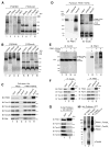
PINK1 complex is associated with components of the TOM machinery. (A) Radiolabeled proteins were imported into isolated HeLa mitochondria in the presence ([35S]-PINK1) or absence ([35S]-Tom40) of 1 μM CCCP for 60 min. Samples were solubilized in 1% digitonin buffer and complexes were immunodepleted using indicated antibodies or beads alone as a control followed by BN-PAGE analysis. (B) Radiolabeled proteins were imported into isolated HeLa mitochondria in the presence ([35S]-PINK1) or absence ([35S]-Tom40) of 1 μM CCCP for 60 min. Samples were solubilized in 1% digitonin buffer followed by the addition of antibodies as indicated, and subjected to BN-PAGE. (C) Mock transfected and PINK1-V5/His stably transfected HeLa cells were treated with 20 μM CCCP for 3 h followed by mitochondrial isolation and immunocapture using α-His antibodies coupled to beads. Bound proteins were eluted with 6xHis peptides and various fractions as indicated were subjected to SDS-PAGE followed by immunoblotting using antibodies as indicated. *, non-specific band. (D) Samples treated as in (C) were analyzed using BN-PAGE and immunoblotting with α-PINK1 (left panel) and α-Tom20 (right panel) antibodies. (E) HeLa cells were either mock transfected or transfected with PINK1-V5/His and treated with or without 20 μM CCCP for 3 h before solubilization in 1% digitonin buffer followed by BN-PAGE and immunoblotting using α-Tom22 (left panel) or α-PINK1 (right panels) antibodies. (F) HEK293 cells were treated with 20 μM CCCP for 3h and then harvested and lysed in 1% digitonin containing buffer. Clarified lysates were used for immunopreciptation using α-Tom20 (left panel) or α-PINK1 (right panel) antibody coupled beads. Input, unbound and bound fractions were subjected to SDS-PAGE and immunoblotted using antibodies against PINK1, Parkin, Tom40 and Tom20. (G) Immunocaptured PINK1-V5/His complex as in (C) was incubated in 0.1 mM dithiobis[succinimidyl propionate] for 20 min on ice. After crosslinking, samples were incubated in 1% SDS containing buffer for 5 min at 95° and then subjected to immunoprecipitation using α-His beads. Crosslinker was cleaved with 100 mM DTT before SDS-PAGE and immunoblotting with antibodies as indicated. (H) Samples were treated as in (G) and subjected to SDS-PAGE in the absence of DTT followed by immunoblotting using α-PINK1 and α-Tom20 antibodies. Radiolabeled proteins were detected by phosphorimage analysis. See also Figure S2.
Figure 4
Figure 4
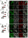
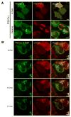
Ectopically localized PINK1 can recruit Parkin to mitochondria, peroxisomes and lysosomes. (A, C, E) Schematic diagrams of organelle specific heterodimerization using FRB-Fis1 for mitochondria (A), PMP34-FRB for peroxisomes (C), and LAMP1-FRB for lysosomes (E) are illustrated. Treatment of cells with rapalog induces the heterodimerization of FRB and FKBP causing cytosolic PINK1Δ110-YFP-FKBP to attach to the organelle specified by the FRBs. (B, D, F) HeLa cells were transfected with PINK1Δ110-YFP-FKBP, CFP-Parkin, one of the organelle specific FRBs and an organelle _targeted fluorescent marker protein: (RFP-SKL for peroxisomes (D) LAMP1-cherry for lysosomes (F)). For mitochondria, cells were stained with MitoTracker Red before rapalog treatment (B). After 48 h of transfection, cells were treated with or without rapalog for 2 h and imaged by confocal microscopy. (D, F) The third rows are enlarged images of the white boxes in the second rows. White bar, 10 μm. See also Figures S3 and S4.
Figure 5
Figure 5
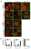
Parkin recruited by ectopic PINK1 can induce autophagy of mitochondria and peroxisomes. (A) HeLa cells were transfected with PINK1Δ110-YFP-FKBP and cherry-Parkin together with one of the organelle specific FRBs (FRB-Fis1 (left column), PMP34-FRB (middle column) and LAMP1-FRB (right column)) for 24 h. After 48 h of treatment with (lower 3 rows) or without (top row) rapalog, cells were fixed, immunostained for organelle specific proteins (pyruvate dehydrogenase subunit E1α (PDH) for mitochondria (left column), catalase for peroxisomes (middle column) and LAMP-2 for lysosomes (right column)) and imaged with confocal microscopy. In rapalog treated cells, representative images of cells showing three different responses (normal, reduced and trace or none) of organelle mass are shown. White boxes on the right bottom corner show only the organelle markers from the dashed box to clearly show the change of organelle mass. White bar, 10 μm. (B) Cells having the indicated amount of each organelle in (A) were counted. To ensure the co-expression of the three constructs (PINK1-FKBP, Parkin and FRB) only the cells showing an abundant expression of mCherry-Parkin were counted. The graphs represent means ± SEM of counts in >100 cell per condition in three independent experiments and analyzed with 2 way ANOVA. ***; P<0.001, **; P<0.01. See also Figure S5.
Figure 6
Figure 6
PINK1 not localized to membranes does not interact with Parkin. (A) Mitochondrial Parkin does not recruit cytosolic PINK1 to mitochondria. HeLa cells were transfected with PINK1Δ110-YFP, mCherry-FKBP-Parkin and FRB-Fis1 and treated with or without rapalog for 2 h. The live cells were imaged by confocal microscopy. (B) Delayed Parkin translocation following the translocation of cytosolic PINK1-FKBP induced by heterodimerization. HeLa cells were transfected with PINK1Δ110-YFP-FKBP, mCherry-Parkin and FRB-Fis1 for 48 h, treated with rapalog and applied for confocal live cell imaging over 17 min. The first image was taken after 2 min of rapalog treatment, and subsequent images were taken after every 5 min. White bar, 10 μm. See also Figure S6.
Figure 7
Figure 7
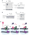
PINK1 and Parkin after CCCP washout. (A) HeLa cells were treated with either DMSO or CCCP for 3 h before CCCP washout for increasing times as indicated. Cells were lysed in 1% digitonin buffer (BN-PAGE; left panel) or SDS sample buffer (SDS-PAGE; right panel) and immunoblotted using α-PINK1 and α-VDAC1 antibodies. (B) PARL−/− MEFs transfected with PINK1-V5/His were treated as in (A). Cells were lysed in SDS sample buffer and immunoblotted using α-PINK1 and α-VDAC1 antibodies. (C) HeLa cells were treated as in (A) and mitochondrial fractions were subjected to SDS-PAGE and immunoblotting using α-Parkin, α-PINK1 and α-Tim23 antibodies. W/O: washout, FL: full-length, ΔMTS: mitochondrial _targeting sequence cleaved. (D) Model for PINK1 regulation. See discussion for description. See also Figure S7.

Similar articles

Cited by

References

    1. Abe Y, Shodai T, Muto T, Mihara K, Torii H, Nishikawa S, Endo T, Kohda D. Structural basis of presequence recognition by the mitochondrial protein import receptor Tom20. Cell. 2000;100:551–560. - PubMed
    1. Ahting U, Thieffry M, Engelhardt H, Hegerl R, Neupert W, Nussberger S. Tom40, the pore-forming component of the protein-conducting TOM channel in the outer membrane of mitochondria. J Cell Biol. 2001;153:1151–1160. - PMC - PubMed
    1. Beilina A, Van Der Brug M, Ahmad R, Kesavapany S, Miller DW, Petsko GA, Cookson MR. Mutations in PTEN-induced putative kinase 1 associated with recessive parkinsonism have differential effects on protein stability. Proc Natl Acad Sci USA. 2005;102:5703–5708. - PMC - PubMed
    1. Belshaw PJ, Ho SN, Crabtree GR, Schreiber SL. Controlling protein association and subcellular localization with a synthetic ligand that induces heterodimerization of proteins. Proc Natl Acad Sci USA. 1996;93:4604–4607. - PMC - PubMed
    1. Brix J, Dietmeier K, Pfanner N. Differential recognition of preproteins by the purified cytosolic domains of the mitochondrial import receptors Tom20, Tom22, and Tom70. J Biol Chem. 1997;272:20730–20735. - PubMed

Publication types

Substances

LinkOut - more resources

  NODES
Association 3
twitter 2