Review
. 2012 Feb 20;196(4):395-406.
doi: 10.1083/jcb.201102147.

The extracellular matrix: a dynamic niche in cancer progression

Affiliations
Review

The extracellular matrix: a dynamic niche in cancer progression

Pengfei Lu et al. J Cell Biol. .

Abstract

The local microenvironment, or niche, of a cancer cell plays important roles in cancer development. A major component of the niche is the extracellular matrix (ECM), a complex network of macromolecules with distinctive physical, biochemical, and biomechanical properties. Although tightly controlled during embryonic development and organ homeostasis, the ECM is commonly deregulated and becomes disorganized in diseases such as cancer. Abnormal ECM affects cancer progression by directly promoting cellular transformation and metastasis. Importantly, however, ECM anomalies also deregulate behavior of stromal cells, facilitate tumor-associated angiogenesis and inflammation, and thus lead to generation of a tumorigenic microenvironment. Understanding how ECM composition and topography are maintained and how their deregulation influences cancer progression may help develop new therapeutic interventions by _targeting the tumor niche.


Figures

Figure 1.
Figure 1.
Mechanisms of ECM function. The versatile functions of the ECM depend on its diverse physical, biochemical, and biomechanical properties. Anchorage to the basement membrane is essential for various biological processes, including asymmetric cell division in stem cell biology and maintenance of tissue polarity (stage 1). Depending on contexts, the ECM may serve to block or facilitate cell migration (stages 2 and 3). In addition, by binding to growth factor signaling molecules and preventing their otherwise free diffusion, the ECM acts as a sink for these signals and helps shape a concentration gradient (stage 4). Certain ECM components, including heparan sulfate proteoglycans and the hyaluronic acid receptor CD44, can selectively bind to different growth factors and function as a signal coreceptor (stage 5) or a presenter (stage 6) and help determine the direction of cell–cell communication (Lu et al., 2011). The ECM also direct signals to the cell by using its endogenous growth factor domains (not depicted) or functional fragment derivatives after being processed by proteases such as MMPs (stage 7). Finally, cells directly sense the biomechanical properties of the ECM, including its stiffness, and change a wide variety of behaviors accordingly (stage 8).
Figure 2.
Figure 2.
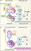
ECM is an essential component of normal and cancer stem cell niche. The ECM plays multiple roles in maintaining stem cell properties. (A) ECM anchorage restricts stem cells in the niche and thus allows them to be exposed to paracrine (stage 1) and cell–cell contact signals (not depicted) that are essential for maintaining stem cell properties. Anchorage is also important for orienting the mitotic spindle and makes it possible for stem cells to undergo asymmetric cell division (stage 2), which is essential for stem cell self-renewal and generation of daughter cells that are destined to undergo cell differentiation. The exact mechanism whereby ECM anchorage controls asymmetric cell division remains unclear, although one possibility is to allow cytoplasmic cell fate determinants to be differentially distributed between the daughter cells. The ECM also maintains stem cell properties via its many other features including its biomechanical properties such as ECM stiffness that affects cell fate determination (stage 3). (B) In the presence of abnormal ECM (pink) or loss of ECM contact, stem cell properties fail to be maintained and undergo symmetric cell division instead, leading to an overexpansion of the (cancer) stem cell pool. Abnormal changes of the ECM can also disrupt the cellular differentiation process, resulting in loss of differentiation and an increase of stem/progenitor cells.
Figure 3.
Figure 3.
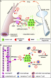
ECM role in tumor angiogenesis, lymphangiogenesis, and inflammation. (A) Angiogenesis and lymphangiogenesis depend on the ECM. Tumor cells produce various components, including VEGF and angiogenic and antiangiogenic ECM fragments, to regulate blood vessel formation (stage 1). During branch initiation, endothelial cells secrete proteases to break down the basement membrane to grow out (stage 2). The outgrowth process of endothelial branching is propelled by at least two groups of cells: tip cells, which lead the migration toward the angiogenic chemoattractant source, and stalk cells, which depend on the ECM and its derivatives to survive and proliferate to provide building blocks for vessel formation (stage 3). Additionally, ECM components participate in cell migration and other aspects of tubulogenesis of blood vessels. Although details remain unclear, lymphangiogenesis depends on the ECM and, together with angiogenesis, provides routes for cancer cell metastasis and immune cell infiltration. (B) The ECM plays multiple roles in tumor inflammation. In addition to promoting survival and proliferation (not depicted), ECM components function as a chemoattractant to immune cells (stage a). The exact details of how immune cells including neutrophil transmigrate endothelial basement membrane are not clear, though it seems the ECM plays both positive and negative roles in the process. Macrophage activation depends on the ECM to release its potent cytokine signals and protease content (stage b). Further, immune cell differentiation, including maturation of T helper cells, requires participation of ECM components (stage c).
Figure 4.
Figure 4.
Abnormal ECM promotes cancer progression. (A) ECM remodeling is tightly controlled to ensure organ homeostasis and functions. Normal ECM dynamics are essential for maintaining tissue integrity and keep rare tumor-prone cells, together with resident fibroblasts, eosinophils, macrophages, and other stromal cells, in check by maintaining an overall healthy microenvironment. (B) With age or under pathological conditions, tissues can enter a series of tumorigenic events. One of the earlier events is the generation of activated fibroblasts or CAFs (stage 1), which contributes to abnormal ECM buildup and deregulated expression of ECM remodeling enzymes (stage 2). Abnormal ECM has profound impacts on surrounding cells, including epithelial, endothelial, and immune cells and other stromal cell types. Deregulated ECM promotes epithelial cellular transformation and hyperplasia (stage 3). (C) In late-stage tumors, immune cells are often recruited to the tumor site to promote cancer progression (stage 4). In addition, deregulated ECM affects various aspects of vascular biology and promotes tumor-associated angiogenesis (stage 5). Creation of a leaky tumor vasculature in turn facilitates tumor cell invasion and metastasis to distant sites (stage 6). (D) At distant sites, cancer cells leave the circulation and take hold of the local tissue. Together with local stromal cells, cancer cells express ECM remodeling enzymes and create a local metastatic niche. Abnormal niche ECM promotes extravasation, survival, and proliferation of cancer cells (stage 7). At later stages when cancer cells awake from dormancy, abnormal ECM turns on the angiogenic switch (stage 8), presumably using a mechanism similar to that used at the primary site (stage 5), and promotes the rapid growth of cancer cells and an expansion of micrometastasis to macrometastasis.

Similar articles

Cited by

References

    1. Adair-Kirk T.L., Senior R.M. 2008. Fragments of extracellular matrix as mediators of inflammation. Int. J. Biochem. Cell Biol. 40:1101–1110 10.1016/j.biocel.2007.12.005 - DOI - PMC - PubMed
    1. Adler B., Ashkar S., Cantor H., Weber G.F. 2001. Costimulation by extracellular matrix proteins determines the response to TCR ligation. Cell. Immunol. 210:30–40 10.1006/cimm.2001.1800 - DOI - PubMed
    1. Aggarwal S., Pittenger M.F. 2005. Human mesenchymal stem cells modulate allogeneic immune cell responses. Blood. 105:1815–1822 10.1182/blood-2004-04-1559 - DOI - PubMed
    1. Aitken K.J., Bägli D.J. 2009. The bladder extracellular matrix. Part I: Architecture, development and disease. Nat Rev Urol. 6:596–611 10.1038/nrurol.2009.201 - DOI - PubMed
    1. Akhtar N., Marlow R., Lambert E., Schatzmann F., Lowe E.T., Cheung J., Katz E., Li W., Wu C., Dedhar S., et al. 2009. Molecular dissection of integrin signalling proteins in the control of mammary epithelial development and differentiation. Development. 136:1019–1027 10.1242/dev.028423 - DOI - PMC - PubMed

Publication types

  NODES
Association 1
chat 1
twitter 2