Crystal structure of TDRD3 and methyl-arginine binding characterization of TDRD3, SMN and SPF30
- PMID: 22363433
- PMCID: PMC3281842
- DOI: 10.1371/journal.pone.0030375
Crystal structure of TDRD3 and methyl-arginine binding characterization of TDRD3, SMN and SPF30
Abstract
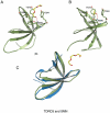
SMN (Survival motor neuron protein) was characterized as a dimethyl-arginine binding protein over ten years ago. TDRD3 (Tudor domain-containing protein 3) and SPF30 (Splicing factor 30 kDa) were found to bind to various methyl-arginine proteins including Sm proteins as well later on. Recently, TDRD3 was shown to be a transcriptional coactivator, and its transcriptional activity is dependent on its ability to bind arginine-methylated histone marks. In this study, we systematically characterized the binding specificity and affinity of the Tudor domains of these three proteins quantitatively. Our results show that TDRD3 preferentially recognizes asymmetrical dimethylated arginine mark, and SMN is a very promiscuous effector molecule, which recognizes different arginine containing sequence motifs and preferentially binds symmetrical dimethylated arginine. SPF30 is the weakest methyl-arginine binder, which only binds the GAR motif sequences in our library. In addition, we also reported high-resolution crystal structures of the Tudor domain of TDRD3 in complex with two small molecules, which occupy the aromatic cage of TDRD3.
Conflict of interest statement
Figures


Similar articles
-
Structural basis for dimethylarginine recognition by the Tudor domains of human SMN and SPF30 proteins.Nat Struct Mol Biol. 2011 Nov 20;18(12):1414-20. doi: 10.1038/nsmb.2185. Nat Struct Mol Biol. 2011. PMID: 22101937
-
Structural plasticity of the TDRD3 Tudor domain probed by a fragment screening hit.FEBS J. 2018 Jun;285(11):2091-2103. doi: 10.1111/febs.14469. Epub 2018 Apr 24. FEBS J. 2018. PMID: 29645362
-
Tudor domains bind symmetrical dimethylated arginines.J Biol Chem. 2005 Aug 5;280(31):28476-83. doi: 10.1074/jbc.M414328200. Epub 2005 Jun 6. J Biol Chem. 2005. PMID: 15955813
-
Tudor domain-containing proteins of Drosophila melanogaster.Dev Growth Differ. 2012 Jan;54(1):32-43. doi: 10.1111/j.1440-169x.2011.01308.x. Dev Growth Differ. 2012. PMID: 23741747 Review.
-
Structure and function of eTudor domain containing TDRD proteins.Crit Rev Biochem Mol Biol. 2019 Apr;54(2):119-132. doi: 10.1080/10409238.2019.1603199. Epub 2019 May 3. Crit Rev Biochem Mol Biol. 2019. PMID: 31046474 Review.
Cited by
-
Site-specific and regiospecific installation of methylarginine analogues into recombinant histones and insights into effector protein binding.J Am Chem Soc. 2013 Feb 27;135(8):2879-82. doi: 10.1021/ja3108214. Epub 2013 Feb 19. J Am Chem Soc. 2013. PMID: 23398247 Free PMC article.
-
Molecular basis underlying histone H3 lysine-arginine methylation pattern readout by Spin/Ssty repeats of Spindlin1.Genes Dev. 2014 Mar 15;28(6):622-36. doi: 10.1101/gad.233239.113. Epub 2014 Mar 3. Genes Dev. 2014. PMID: 24589551 Free PMC article.
-
Chromatin dependencies in cancer and inflammation.Nat Rev Mol Cell Biol. 2018 Apr;19(4):245-261. doi: 10.1038/nrm.2017.113. Epub 2017 Nov 29. Nat Rev Mol Cell Biol. 2018. PMID: 29184195 Review.
-
A small molecule antagonist of SMN disrupts the interaction between SMN and RNAP II.Nat Commun. 2022 Sep 16;13(1):5453. doi: 10.1038/s41467-022-33229-5. Nat Commun. 2022. PMID: 36114190 Free PMC article.
-
Structural insights into the sequence-specific recognition of Piwi by Drosophila Papi.Proc Natl Acad Sci U S A. 2018 Mar 27;115(13):3374-3379. doi: 10.1073/pnas.1717116115. Epub 2018 Mar 12. Proc Natl Acad Sci U S A. 2018. PMID: 29531043 Free PMC article.
References
-
- Bedford MT, Richard S. Arginine methylation an emerging regulator of protein function. Mol Cell. 2005;18:263–272. - PubMed
-
- Friesen WJ, Massenet S, Paushkin S, Wyce A, Dreyfuss G. SMN, the product of the spinal muscular atrophy gene, binds preferentially to dimethylarginine-containing protein _targets. Mol Cell. 2001;7:1111–1117. - PubMed
Publication types
MeSH terms
Substances
Grants and funding
LinkOut - more resources
Full Text Sources

