. 2012 Jun;86(12):6537-45.
doi: 10.1128/JVI.00094-12. Epub 2012 Apr 11.

Simultaneous treatment of human bronchial epithelial cells with serine and cysteine protease inhibitors prevents severe acute respiratory syndrome coronavirus entry

Affiliations

Simultaneous treatment of human bronchial epithelial cells with serine and cysteine protease inhibitors prevents severe acute respiratory syndrome coronavirus entry

Miyuki Kawase et al. J Virol. 2012 Jun.

Abstract

The type II transmembrane protease TMPRSS2 activates the spike (S) protein of severe acute respiratory syndrome coronavirus (SARS-CoV) on the cell surface following receptor binding during viral entry into cells. In the absence of TMPRSS2, SARS-CoV achieves cell entry via an endosomal pathway in which cathepsin L may play an important role, i.e., the activation of spike protein fusogenicity. This study shows that a commercial serine protease inhibitor (camostat) partially blocked infection by SARS-CoV and human coronavirus NL63 (HCoV-NL63) in HeLa cells expressing the receptor angiotensin-converting enzyme 2 (ACE2) and TMPRSS2. Simultaneous treatment of the cells with camostat and EST [(23,25)trans-epoxysuccinyl-L-leucylamindo-3-methylbutane ethyl ester], a cathepsin inhibitor, efficiently prevented both cell entry and the multistep growth of SARS-CoV in human Calu-3 airway epithelial cells. This efficient inhibition could be attributed to the dual blockade of entry from the cell surface and through the endosomal pathway. These observations suggest camostat as a candidate antiviral drug to prevent or depress TMPRSS2-dependent infection by SARS-CoV.


Figures

Fig 1
Fig 1
Enhancement of protease-mediated viral entry into cells. (A) Pseudotyped viruses bearing SARS-S, NL63-S, or VSV-G were incubated with serially diluted trypsin for 5 min at 37°C, followed by inoculation onto HeLa-ACE2 cells for 30 min on ice. (B) Pseudotyped viruses were first adsorbed onto HeLa-ACE2 cells for 30 min on ice, followed by the addition of serial dilutions of trypsin to cells and incubation for a further 5 min at 37°C. The medium was then changed, and the cells were incubated for 20 h at 37°C. Infectivity was quantified by counting GFP-positive cells. The results are presented as the percentage of infected cells (at least 50 GFP-positive cells were counted). Error bars indicate the standard deviations (SD) of the means from four independent wells.
Fig 2
Fig 2
Effect of TMPRSS2 on virus entry into cells. (A) Pseudotyped viruses were inoculated onto HeLa-ACE2 (white bars) and HeLa-ACE2-TMPRSS2 (black bars) cells. Infectivity was quantified by counting GFP-positive cells 20 h after viral inoculation. The results are presented as the percentage of infected HeLa-ACE2 cells (at least 50 GFP-positive cells were counted). Error bars indicate the SD of the means from eight independent samples. (B). Authentic SARS-CoV and HCoV-NL63 were inoculated onto HeLa-ACE2 (white bars) and HeLa-ACE2-TMPRSS2 (black bars) cells. Infectivity was quantified by real-time PCR to measure the viral mRNA9 of these viruses. Error bars indicate the SD of the means from four independent samples.
Fig 3
Fig 3
Effects of serine protease inhibitors on viral entry and syncytium formation. (A) HeLa-ACE2 (white bars) and HeLa-ACE2-TMPRSS2 (black bars) cells were infected with pseudotype viruses bearing SARS-S, NL63-S, or VSV-G in the presence of 10 μM benzamidine (Benz), 10 μM aprotinin (Apro), 10 μM gabexate (Gabex), 10 μM camostat (Camo), or 50 μM TLCK. (B) Concentration-dependent effects of inhibitors in HeLa-ACE2 (triangles) or HeLa-ACE2-TMPRSS2 (circles) cells infected with pseudotyped viruses bearing SARS-S, NL63-S, or VSV-G in the presence of serially diluted camostat or gabexate. Infectivity was quantified by counting GFP-positive cells 20 h after viral inoculation. The results are presented as a percentage of infection (at least 200 GFP-positive cells were counted for untreated [No] cells), and error bars indicate the SD of the means from four independent wells.
Fig 4
Fig 4
Inhibition of syncytium formation by camostat. (A) Plasmids encoding SARS-S or NL63-S were transfected with plasmid encoding enhanced green fluorescent protein (EGFP) in HeLa-ACE2-TMPRSS2 cells, and incubated in the presence or absence of 10 μM camostat. After 20 h, the cells were fixed with 4% formalin and stained with DAPI. (B) The sizes of syncytia in the absence or presence of 0.1, 1, 10, or 100 μM camostat were quantified by counting the number of nuclei in the fused cells. Error bars indicate the SD of the means from 10 independent syncytia. ND, not determined.
Fig 5
Fig 5
Effects of cathepsin inhibitors or endosome-tropic inhibitors on viral entry. HeLa-ACE2 (white bars) and HeLa-ACE2-TMPRSS2 (black bars) cells were infected with pseudotyped viruses bearing SARS-S, NL63-S, or VSV-G in the presence of 100 nM bafilomycin A1 (Baf), 10 μM EST, 50 μM MDL28170 (MDL), 5 μM cathepsin L inhibitor III (LIII), 50 μM leupeptin (Leu), or 1 μM cytochalasin D (CytD). Infectivity was quantified by counting GFP-positive cells 20 h after viral inoculation. The results are presented as a percentage of infection (at least 200 GFP-positive cells were counted for untreated [No] cells), and error bars indicate the SD of the means from four independent wells.
Fig 6
Fig 6
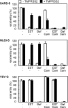
Inhibition of viral entry by simultaneous treatment with serine and cysteine protease inhibitors. HeLa-ACE2 (white bars) and HeLa-ACE2-TMPRSS2 (black bars) cells were infected with pseudotyped viruses bearing SARS-S, NL63-S, or VSV-G in the presence of 10 μM EST, 10 μM camostat (Cam), 100 nM bafilomycin (Baf), both EST and Cam, or both bafilomycin and camostat. Infectivity was quantified by counting GFP-positive cells 20 h after viral inoculation. The results are presented as a percentage of infection (at least 200 GFP-positive cells were counted for untreated cells), and error bars indicate the SD of the means from four independent wells.
Fig 7
Fig 7
Cell entry kinetics of pseudotyped viruses. Pseudotyped viruses bearing SARS-S, NL63-S, or VSV-G were inoculated onto HeLa-ACE2 (triangles) and HeLa-ACE2-TMPRSS2 (circles) cells and treated with both 10 μM EST and 10 μM camostat to stop viral entry at the indicated time points. Infectivity was quantified by counting GFP-positive cells 20 h after viral inoculation. The results are presented as a percentage of infection (at least 500 GFP-positive cells were counted for untreated cells), and error bars indicate the SD of the means from four independent wells.
Fig 8
Fig 8
Comparison of transcripts in human lung and Calu-3 cells. The amounts of ACE2, TMPRSS2 (TM2), and HAT mRNA in human lungs or Calu-3 cells were measured using real-time PCR as described in Materials and Methods. Error bars indicate the SD of the means from six independent samples.
Fig 9
Fig 9
Effect of TMPRSS2 and HAT siRNA-mediated knockdown on SARS-CoV entry into Calu-3 cells. (A) The cells were transiently transfected with siRNAs _targeting TMPRSS2 (TM2-_target) or HAT (HAT-_target) or a non_targeting control (“Non-_target”). Nontransfected cells served as the negative control (−). Four days after transfection, cellular RNA was isolated, and ACE2, TMPRSS2, and HAT mRNAs were measured by real-time PCR. (B) Calu-3 cells were infected with SARS-CoV 4 days after transfection of a non_targeting siRNA or nontransfected control (−). Cellular RNA was isolated 5 h after infection, and infectivity was quantified by real-time PCR. (C) Four days after the above-described transfection with siRNA, Calu-3 cells were infected with SARS-CoV in the presence or absence of 10 μM EST. Cellular RNA was isolated 5 h after infection, and infectivity was quantified by real-time PCR measurement of the amount of viral mRNA9. Error bars indicate the SD of the means from six independent samples.
Fig 10
Fig 10
Inhibition of viral entry and multistep growth of SARS-CoV in Calu-3 cells after simultaneous treatment with inhibitors. (A) Calu-3 cells were infected with SARS-CoV at an MOI of 10 in the presence of 10 μM camostat (Cam), 10 μM EST, or both. Five hours later, cellular RNA was isolated and infectivity was quantified by real-time PCR. (B) The mRNA levels for a housekeeping gene (GAPDH) and for the virus receptor (ACE2) were measured to evaluate the nontoxicity of camostat and EST in the same Calu-3 cells for which the infectivity results are shown in panel A. (C) Calu-3 cells were infected with SARS-CoV at an MOI of 0.01 for 1 h at 37°C, and then the cells were incubated in the presence of 10 μM camostat, 10 μM EST, or both. Nontreated cells served as the negative control. Cellular RNA was isolated on the indicated days. (D) GAPDH and ACE2 mRNA levels were measured to evaluate the nontoxicity of camostat and EST in the samples at 6 days postinfection (shown in panel C). Infectivity was quantified by real-time PCR measurement of the amount of viral mRNA9. Error bars indicate the SD of the means from six independent samples.

Similar articles

Cited by

References

    1. Belouzard S, Chu VC, Whittaker GR. 2009. Activation of the SARS coronavirus spike protein via sequential proteolytic cleavage at two distinct sites. Proc. Natl. Acad. Sci. U. S. A. 106:5871–5876 - PMC - PubMed
    1. Belouzard S, Madu I, Whittaker GR. 2010. Elastase-mediated activation of the severe acute respiratory syndrome coronavirus spike protein at discrete sites within the S2 domain. J. Biol. Chem. 285:22758–22763 - PMC - PubMed
    1. Bertram S, et al. 2010. TMPRSS2 and TMPRSS4 facilitate trypsin-independent spread of influenza virus in Caco-2 cells. J. Virol. 84:10016–10025 - PMC - PubMed
    1. Bertram S, et al. 2011. Cleavage and activation of the severe acute respiratory syndrome coronavirus spike protein by human airway trypsin-like protease. J. Virol. 85:13363–13372 - PMC - PubMed
    1. Bertram S, Glowacka I, Steffen I, Kühl A, Pöhlmann S. 2010. Novel insights into proteolytic cleavage of influenza virus hemagglutinin. Rev. Med. Virol. 20:298–310 - PMC - PubMed

Publication types

MeSH terms

Substances

  NODES
INTERN 1
twitter 2