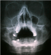
Accidental displacement and migration of endosseous implants into adjacent craniofacial structures: a review and update
- PMID: 22549685
- PMCID: PMC3482520
- DOI: 10.4317/medoral.18032
Accidental displacement and migration of endosseous implants into adjacent craniofacial structures: a review and update
Abstract
Objectives: Accidental displacement of endosseous implants into the maxillary sinus is an unusual but potential complication in implantology procedures due to the special features of the posterior aspect of the maxillary bone; there is also a possibility of migration throughout the upper paranasal sinuses and adjacent structures. The aim of this paper is to review the published literature about accidental displacement and migration of dental implants into the maxillary sinus and other adjacent structures.
Study design: A review has been done based on a search in the main on-line medical databases looking for papers about migration of dental implants published in major oral surgery, periodontal, dental implant and ear-nose-throat journals, using the keywords "implant," "migration," "complication," "foreign body" and "sinus."
Results: 24 articles showing displacement or migration to maxillary, ethmoid and sphenoid sinuses, orbit and cranial fossae, with different degrees of associated symptoms, were identified. Techniques found to solve these clinical issues include Cadwell-Luc approach, transoral endoscopy approach via canine fossae and transnasal functional endoscopy surgery.
Conclusion: Before removing the foreign body, a correct diagnosis should be done in order to evaluate the functional status of the ostiomeatal complex and the degree of affectation of paranasal sinuses and other involved structures, determining the size and the exact location of the foreign body. After a complete diagnosis, an indicated procedure for every case would be decided.
Figures


Similar articles
-
Displacement of a dental implant into the maxillary sinus: case series.Minerva Stomatol. 2010 Jan-Feb;59(1-2):45-54. Minerva Stomatol. 2010. PMID: 20212409 English, Italian.
-
The management of complications following displacement of oral implants in the paranasal sinuses: a multicenter clinical report and proposed treatment protocols.Int J Oral Maxillofac Surg. 2009 Dec;38(12):1273-8. doi: 10.1016/j.ijom.2009.09.001. Epub 2009 Sep 24. Int J Oral Maxillofac Surg. 2009. PMID: 19781911 Clinical Trial.
-
Removal of dental implant displaced into maxillary sinus by combination of endoscopically assisted and bone repositioning techniques: a case report.J Med Case Rep. 2016 Jan 12;10:1. doi: 10.1186/s13256-015-0787-1. J Med Case Rep. 2016. PMID: 26758705 Free PMC article.
-
Implants Displaced Into the Maxillary Sinus: A Systematic Review.Implant Dent. 2016 Aug;25(4):547-51. doi: 10.1097/ID.0000000000000408. Implant Dent. 2016. PMID: 26974033 Review.
-
Effect of maxillary sinus augmentation on the survival of endosseous dental implants. A systematic review.Ann Periodontol. 2003 Dec;8(1):328-43. doi: 10.1902/annals.2003.8.1.328. Ann Periodontol. 2003. PMID: 14971260 Review.
Cited by
-
Extraordinary sneeze: Spontaneous transmaxillary-transnasal discharge of a migrated dental implant.World J Clin Cases. 2016 Aug 16;4(8):229-32. doi: 10.12998/wjcc.v4.i8.229. World J Clin Cases. 2016. PMID: 27574611 Free PMC article.
-
Incisive dental implant migration into the nasal septum.BMJ Case Rep. 2019 Jul 27;12(7):e228325. doi: 10.1136/bcr-2018-228325. BMJ Case Rep. 2019. PMID: 31352375 Free PMC article.
-
Unexpected foreign body induced refractory maxillary sinusitis.Clin Case Rep. 2021 Feb 20;9(4):2185-2188. doi: 10.1002/ccr3.3976. eCollection 2021 Apr. Clin Case Rep. 2021. PMID: 33936660 Free PMC article.
-
Treatment of dental implant displacement into the maxillary sinus.Maxillofac Plast Reconstr Surg. 2017 Nov 25;39(1):35. doi: 10.1186/s40902-017-0133-1. eCollection 2017 Dec. Maxillofac Plast Reconstr Surg. 2017. PMID: 29204419 Free PMC article. Review.
-
Nasal septal foreign body as a complication of dental root canal therapy: A case report.World J Clin Cases. 2021 Jan 26;9(3):690-696. doi: 10.12998/wjcc.v9.i3.690. World J Clin Cases. 2021. PMID: 33553410 Free PMC article.
References
-
- Leonhardt A, Gröndahl K, Bergstrom C, Lekhölm U. Long-term follow-up of osseointegrated titanium implants using clinical, radiographic and microbiological parameters. Clin Oral Implants Res. 2002;13:127–32. - PubMed
-
- Atwood DA. Reduction of residual ridges: a major oral disease entity. J Prosthet Dent. 1971;26:266–79. - PubMed
-
- McAllister BS, Haghighat K. Bone augmentation techniques. J Periodontol. 2007;78:377–96. - PubMed
-
- Friberg B, Jemt T, Lekholm U. Early failures in 4,641 consecutively placed Brånemark dental implants: a study from stage 1 surgery to the connection of completed prostheses. Int J Oral Maxillofac Implants. 1991;6:142–6. - PubMed
-
- Cawood JI, Howell RA. A classification of the edentulous jaws. Int J Oral Maxillofac Surg. 1988;17:232–6. - PubMed
Publication types
MeSH terms
Substances
LinkOut - more resources
Full Text Sources

