. 2012 Jul 15;189(2):1014-23.
doi: 10.4049/jimmunol.1200857. Epub 2012 Jun 15.

Molecular mechanisms responsible for the selective and low-grade induction of proinflammatory mediators in murine macrophages by lipopolysaccharide

Affiliations

Molecular mechanisms responsible for the selective and low-grade induction of proinflammatory mediators in murine macrophages by lipopolysaccharide

Urmila Maitra et al. J Immunol. .

Abstract

Low-dose endotoxemia is prevalent in humans with adverse health conditions, and it correlates with the pathogenesis of chronic inflammatory diseases such as atherosclerosis, diabetes, and neurologic inflammation. However, the underlying molecular mechanisms are poorly understood. In this study, we demonstrate that subclinical low-dose LPS skews macrophages into a mild proinflammatory state, through cell surface TLR4, IL-1R-associated kinase-1, and the Toll-interacting protein. Unlike high-dose LPS, low-dose LPS does not induce robust activation of NF-κB, MAPKs, PI3K, or anti-inflammatory mediators. Instead, low-dose LPS induces activating transcription factor 2 through Toll-interacting protein-mediated generation of mitochondrial reactive oxygen species, allowing mild induction of proinflammatory mediators. Low-dose LPS also suppresses PI3K and related negative regulators of inflammatory genes. Our data reveal novel mechanisms responsible for skewed and persistent low-grade inflammation, a cardinal feature of chronic inflammatory diseases.


Figures

Figure 1
Figure 1. Low dose LPS selectively induces mild and prolonged expression of pro-inflammatory mediators
(A) WT BMDM were treated with either a low dose LPS (50 pg/mL) or a high dose LPS (100 ng/mL) for the specified time periods. Total RNA was isolated, and real-time RT-PCR assays were performed to determine the expression levels of pro-inflammatory mediators such as Il-6 and Tnfα. (B) WT BMDM cells were treated with either a low dose LPS (50 pg/mL) or a high dose LPS (200 ng/mL) for the specified time periods. The levels of anti-inflammatory Il-10 and Ccl22 were measured by real-time RT-PCR. The relative transcript levels were standardized against Gapdh levels. Data were representative of at least three independent experiments. * p < 0.05.
Figure 2
Figure 2. Low dose LPS does not induce the classical NFκB and MAPK pathways, nor negative suppressors for inflammatory responses
(A) Low dose LPS fails to induce IκBα degradation. WT BMDM were treated with either a low-dose LPS (50 pg/mL) or a high-dose LPS (100 ng/mL) for the indicated times. The levels of IkBα were determined by Western blot. The same blots were probed with Abs specific for GAPDH as loading controls. (B) Low-dose LPS failed to activate the MAP kinase pathways. Whole cell lysates from WT cells treated with either a low (50pg/mL) or high (200ng/mL) dose LPS were harvested and used to determine the levels of phosphorylated JNK, p38 and ERK. The total levels of JNK, p38 and ERK were used as loading controls. (C) Low-dose LPS fails to induce the negative regulators including MKP-1. Total levels of MKP-1 were determined by Western blot. (D) Low dose LPS reduces IRAK-M. Total levels of IRAK-M from cells treated with either a low or high dose LPS were measured by Western blot. (E) Low dose LPS fails to degrade IRAK-1. IRAK-1 levels were determined from cells treated with either low dose or high dose LPS by Western blot. Levels of GAPDH were used as loading controls in Fig C, D, and e. (F) PI3K pathway is activated by high dose LPS, and suppressed by low dose LPS. Whole cell lysates from cells treated with either low or high dose LPS were harvested and used to determine the levels of phosphorylated Akt. The total levels of Akt were used as the loading control. (G) Nuclear GSK3β is induced by low dose LPS, and reduced by high dose LPS. Nuclear lysates from cells treated with low or high dose LPS were harvested and the levels of GSK3β were determined by Western blot. The levels of Lamin B were used as a loading control. Panels were representatives of three independent experiments. (H) CHIP analysis to detect the binding of RelB to the Il-6 promoter in response to high and low dose LPS. WT BMDM cells were treated with either low or high dose LPS for the indicated time periods. The samples were immunoprecipitated using a RelB specific antibody and analyzed by PCR using primers spanning the promoter region of murine Il-6.
Figure 3
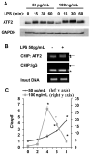
Figure 3. Low dose LPS selectively induces ATF2
(A) Low-dose LPS induces a rapid increase in ATF-2 protein levels in WT BMDM. WT cells were treated with a low (50 pg/mL) or a high dose (100 ng/mL) LPS for the indicated times. Total ATF2 protein levels were determined by Western blot and GAPDH levels were used as loading controls. (B) Low dose LPS recruits ATF2 to the proximal promoter of the pro-inflammatory gene IL-6 in WT BMDMs. BMDM were either untreated or treated with 50 pg/mL LPS for 2 h and then subjected to ChIP assay using an antibody specific to ATF-2 and primers specific to the proximal promoter of IL-6. The same samples were immunoprecipitated using IgG as a non-specific control and input DNA was analyzed as the loading control. Arrow points to the specific amplification product. * denotes a non-specific band. (C) Low-dose LPS induces a late C/ebpδ expression in WT BMDM. WT cells were treated with either a low (50 pg/mL) or a high dose (100 ng/mL) LPS for the indicated times. C/ebpδ message levels were analyzed by real-time RT-PCR. n=3; * p<0.05.
Figure 4
Figure 4. Low dose LPS mediated activation of ATF2 requires IRAK-1 and Tollip
(A) Reduced induction of Il-6 and Tnfα in IRAK-1 and Tollip deficient BMDM cells. WT, IRAK-1 and Tollip deficient BMDM were treated with low dose LPS (50pg/mL) for 4 hrs and the transcript levels of Il-6 and Tnfα were analyzed by real time RT-PCR. (B) Induction of ATF2 in response to low dose LPS depends on IRAK-1 and Tollip. Whole cell lysates were prepared from WT, IRAK-1 and Tollip deficient BMDMs treated with low dose LPS for the indicated time points and analyzed by Western blot. The same blots were probed with GAPDH as loading controls. The adjusted resting levels of ATF2 in each cell type were set as 1, and the ATF2 levels in cells treated with LPS were compared and plotted. n=3; *p<0.05. (C) Low-dose LPS mediated induction of C/ebpδ message is dependent on IRAK-1. WT, IRAK-1 and Tollip deficient BMDM were treated with low dose LPS (50pg/mL) for 6 hrs and the C/ebpδ message levels were analyzed and plotted as shown. (D) Tollip and IRAK- 1 are required for low dose LPS mediated recruitment of ATF2 to the promoter of Il-6. WT, IRAK-1 and Tollip deficient BMDMs were either untreated or treated with 50 pg/mL LPS for 2 h and subjected to ChIP assay using antibodies specific to ATF-2 or C/EBPδ and primers specific to the proximal promoter of Il-6. The same samples were immunoprecipitated using IgG as specificity control and input DNA was analyzed as the loading control. Arrow points to the specific amplification product. * denotes a non-specific band.
Figure 5
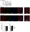
Figure 5. Low dose LPS translocates Tollip to the mitochondria and induces ROS
(A) Mitochondrial localization of Tollip in response to low dose LPS using subcellular fractionation. WT BMDM cells were treated with low dose LPS and mitochondrial protein fractions were prepared. The purity of the mitochondrial fraction was determined using Western blot analysis for mitochondrial resident protein cyclooxygenase IV and cytosolic protein, GAPDH. (B) Mitochondrial localization of Tollip in response to low dose LPS using confocal microscopy. The GFP-Tollip transfected MEF cells were stained with Mito Tracker Red to stain the mitochondria. The nuclei were stained using DAPI (blue). The cells were visualized under laser-scanning confocal microscope. The merged images were magnified and shown on the right panel and the co-localization is indicated with an arrow. (C) Involvement of Tollip in mitochondrial ROS generation. WT, IRAK-1 and Tollip deficient BMDM were stained with mitochondrial ROS selective dye, MitoSOX Red. Cells were then washed with HBSS and treated with low dose LPS for 60 minutes. The cells were also stained with 1 μM Hoescht 34580 for normalization of fluorescent data. Fluorescent intensities were measured and quantified. n=3; * p<0.05.
Figure 6
Figure 6. Mitochondrial ROS contributes to induction of low grade inflammation
(A) The induction of ATF2 by low dose LPS is blocked by ROS inhibitor. WT BMDM cells treated with low dose LPS (50 pg/mL) in the presence or absence of NAC for the indicated time points. Total cellular lysates were analyzed for ATF2 induction in response to low dose LPS. The band intensities were quantitated and the relative fold expressions were plotted. n=3; * p<0.05. (B) Inhibition of low dose LPS induced Il-6 by ROS inhibitor. WT BMDM cells were pretreated with DMSO, NAC or an IKKε inhibitor (IKKin) for 30 min and then treated with low dose LPS (50 pg/mL) for 4 hrs. The cells were harvested and the total RNA was used to detect the transcript levels of Il-6.
Figure 7
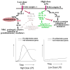
Figure 7. A schematic illustration of potential mechanisms responsible for low grade inflammation induced by low dose LPS
Low dose LPS activates ATF2 through IRAK-1 and Tollip-mediated production of mitochondria ROS. In the meantime, low dose LPS suppresses PI3K, IRAK-M, and other related negative regulators. As a consequence, low dose LPS induces mild, prolonged, and skewed expression of pro-inflammatory mediators. In contrast, high dose LPS potently activated NFκB pathway, as well as the PI3K pathway, and causes robust yet transient expression of pro- and anti-inflammatory mediators.

Similar articles

Cited by

References

    1. Cani PD, Bibiloni R, Knauf C, Waget A, Neyrinck AM, Delzenne NM, Burcelin R. Changes in gut microbiota control metabolic endotoxemia-induced inflammation in high-fat diet-induced obesity and diabetes in mice. Diabetes. 2008;57:1470–1481. - PubMed
    1. Goto T, Eden S, Nordenstam G, Sundh V, Svanborg-Eden C, Mattsby-Baltzer I. Endotoxin levels in sera of elderly individuals. Clin Diagn Lab Immunol. 1994;1:684–688. - PMC - PubMed
    1. Ancuta P, Kamat A, Kunstman KJ, Kim EY, Autissier P, Wurcel A, Zaman T, Stone D, Mefford M, Morgello S, Singer EJ, Wolinsky SM, Gabuzda D. Microbial translocation is associated with increased monocyte activation and dementia in AIDS patients. PLoS One. 2008;3:e2516. - PMC - PubMed
    1. Szeto CC, Kwan BC, Chow KM, Lai KB, Chung KY, Leung CB, Li PK. Endotoxemia is related to systemic inflammation and atherosclerosis in peritoneal dialysis patients. Clin J Am Soc Nephrol. 2008;3:431–436. - PMC - PubMed
    1. Wiedermann CJ, Kiechl S, Dunzendorfer S, Schratzberger P, Egger G, Oberhollenzer F, Willeit J. Association of endotoxemia with carotid atherosclerosis and cardiovascular disease prospective results from the Bruneck Study. J Am Coll Cardiol. 1999;34:1975–1981. - PubMed

Publication types

MeSH terms

Substances

  NODES
Association 1
Note 2
twitter 2