The relationship of topographical memory performance to regional neurodegeneration in Alzheimer's disease
- PMID: 22783190
- PMCID: PMC3389330
- DOI: 10.3389/fnagi.2012.00017
The relationship of topographical memory performance to regional neurodegeneration in Alzheimer's disease
Abstract
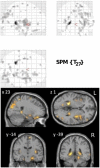
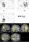
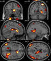
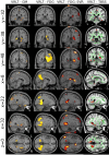
The network activated during normal route learning shares considerable homology with the network of degeneration in the earliest symptomatic stages of Alzheimer's disease (AD). This inspired the virtual route learning test (VRLT) in which patients learn routes in a virtual reality environment. This study investigated the neural basis of VRLT performance in AD to test whether impairment was underpinned by a network or by the widely held explanation of hippocampal degeneration. VRLT score in a mild AD cohort was regressed against gray matter (GM) density and diffusion tensor metrics of white matter (WM) (n = 30), and, cerebral glucose metabolism (n = 26), using a mass univariate approach. GM density and cerebral metabolism were then submitted to a multivariate analysis [support vector regression (SVR)] to examine whether there was a network associated with task performance. Univariate analyses of GM density, metabolism and WM axial diffusion converged on the vicinity of the retrosplenial/posterior cingulate cortex, isthmus and, possibly, hippocampal tail. The multivariate analysis revealed a significant, right hemisphere-predominant, network level correlation with cerebral metabolism; this comprised areas common to both activation in normal route learning and early degeneration in AD (retrosplenial and lateral parietal cortices). It also identified right medio-dorsal thalamus (part of the limbic-diencephalic hypometabolic network of early AD) and right caudate nucleus (activated during normal route learning). These results offer strong evidence that topographical memory impairment in AD relates to damage across a network, in turn offering complimentary lesion evidence to previous studies in healthy volunteers for the neural basis of topographical memory. The results also emphasize that structures beyond the mesial temporal lobe (MTL) contribute to memory impairment in AD-it is too simplistic to view memory impairment in AD as a synonym for hippocampal degeneration.
Keywords: Alzheimer's; MRI; PET; multivariate; retrosplenial cortex; support vector; topographical memory.
Figures


Similar articles
-
Associations between white matter microstructure and amyloid burden in preclinical Alzheimer's disease: A multimodal imaging investigation.Neuroimage Clin. 2014 Feb 19;4:604-14. doi: 10.1016/j.nicl.2014.02.001. eCollection 2014. Neuroimage Clin. 2014. PMID: 24936411 Free PMC article.
-
Diffusion tensor imaging in Alzheimer's disease: insights into the limbic-diencephalic network and methodological considerations.Front Aging Neurosci. 2014 Oct 2;6:266. doi: 10.3389/fnagi.2014.00266. eCollection 2014. Front Aging Neurosci. 2014. PMID: 25324775 Free PMC article. Review.
-
Parallel ICA of FDG-PET and PiB-PET in three conditions with underlying Alzheimer's pathology.Neuroimage Clin. 2014 Mar 19;4:508-16. doi: 10.1016/j.nicl.2014.03.005. eCollection 2014. Neuroimage Clin. 2014. PMID: 24818077 Free PMC article.
-
Absolute diffusivities define the landscape of white matter degeneration in Alzheimer's disease.Brain. 2010 Feb;133(Pt 2):529-39. doi: 10.1093/brain/awp257. Epub 2009 Nov 13. Brain. 2010. PMID: 19914928
-
Diffusion tensor imaging of white matter degeneration in Alzheimer's disease and mild cognitive impairment.Neuroscience. 2014 Sep 12;276:206-15. doi: 10.1016/j.neuroscience.2014.02.017. Epub 2014 Feb 27. Neuroscience. 2014. PMID: 24583036 Review.
Cited by
-
The relationship between vestibular function and topographical memory in older adults.Front Integr Neurosci. 2014 Jun 2;8:46. doi: 10.3389/fnint.2014.00046. eCollection 2014. Front Integr Neurosci. 2014. PMID: 24917795 Free PMC article.
-
Molecular imaging identifies age-related attenuation of acetylcholine in retrosplenial cortex in response to acetylcholinesterase inhibition.Neuropsychopharmacology. 2019 Nov;44(12):2091-2098. doi: 10.1038/s41386-019-0397-5. Epub 2019 Apr 22. Neuropsychopharmacology. 2019. PMID: 31009936 Free PMC article.
-
Behavioral Phenotype in the TgF344-AD Rat Model of Alzheimer's Disease.Front Neurosci. 2020 Jun 16;14:601. doi: 10.3389/fnins.2020.00601. eCollection 2020. Front Neurosci. 2020. PMID: 32612506 Free PMC article.
-
A similarity-based approach to leverage multi-cohort medical data on the diagnosis and prognosis of Alzheimer's disease.Gigascience. 2018 Jul 1;7(7):giy085. doi: 10.1093/gigascience/giy085. Gigascience. 2018. PMID: 30010762 Free PMC article.
-
Tart Cherry Extract and Omega Fatty Acids Reduce Behavioral Deficits, Gliosis, and Amyloid-Beta Deposition in the 5xFAD Mouse Model of Alzheimer's Disease.Brain Sci. 2021 Oct 27;11(11):1423. doi: 10.3390/brainsci11111423. Brain Sci. 2021. PMID: 34827424 Free PMC article.
References
-
- Braak H., Braak E. (1991). Neuropathological stageing of Alzheimer-related changes. Acta Neuropathol. 82, 239–259 - PubMed
Grants and funding
LinkOut - more resources
Full Text Sources

