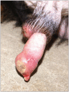
Primary cutaneous undifferentiated round cell tumor with concurrent polymyositis in a dog
- PMID: 23115370
- PMCID: PMC3327596
Primary cutaneous undifferentiated round cell tumor with concurrent polymyositis in a dog
Abstract
A cutaneous poorly differentiated round cell tumor with concurrent, non-suppurative, polymyositis was diagnosed in a hovawart dog. Histochemical staining, immunohistochemistry, and transmission electron microscopy findings suggested that the tumors cells were of myeloid, or possibly natural killer cell origin. The possibility that the concurrent polymyositis may represent a pre-neoplastic or paraneoplastic process is discussed.
Tumeur cutanée primaire indifférenciée à cellules rondes avec polymyosite concomitante chez un chien. Une tumeur cutanée à cellules rondes mal différenciée avec polymyosite non suppurative concomitante a été diagnostiquée chez un chien Hovawart. Les résultats de la coloration histochimique, de l’immunohistochimie et de la microscopie électronique à transmission ont suggéré que les cellules de la tumeur étaient d’origine myéloïde ou possiblement des cellules tueuses naturelles. La possibilité que la polymyosite concomitante puisse représenter un processus néoplasique ou paranéoplasique est examinée.
(Traduit par Isabelle Vallières)
Figures


Similar articles
-
Immunohistochemical and histochemical stains for differentiating canine cutaneous round cell tumors.Vet Pathol. 2005 Jul;42(4):437-45. doi: 10.1354/vp.42-4-437. Vet Pathol. 2005. PMID: 16006603
-
Dilatation of the right atrium in a dog with polymyositis and myocarditis.J Small Anim Pract. 2008 Jun;49(6):302-5. doi: 10.1111/j.1748-5827.2007.00516.x. Epub 2008 Mar 26. J Small Anim Pract. 2008. PMID: 18373536
-
Symptomatic tongue atrophy due to atypical polymyositis in a Pembroke Welsh Corgi.J Vet Med Sci. 2009 Aug;71(8):1063-7. doi: 10.1292/jvms.71.1063. J Vet Med Sci. 2009. PMID: 19721359
-
Review of diagnostic histologic features of cutaneous round cell neoplasms in dogs.J Vet Diagn Invest. 2022 Sep;34(5):769-779. doi: 10.1177/10406387221100209. Epub 2022 Jun 2. J Vet Diagn Invest. 2022. PMID: 35655419 Free PMC article. Review.
-
Canine histiocytic diseases.Compend Contin Educ Vet. 2008 Apr;30(4):202-4, 208-16; quiz 216-17. Compend Contin Educ Vet. 2008. PMID: 18576276 Review.
References
-
- Kaddu S, Beham-Schmid C, Zenahlik P, Kerl H, Cerroni L. CD56+ blastic transformation of chronic myeloid leukemia involving the skin. J Cutan Pathol. 1999;26:497–503. - PubMed
-
- Kaddu S, Zenahlik P, Beham-Schmid C, Kerl H, Cerroni L. Specific cutaneous infiltrates in patients with myelogenous leukemia: A clinicopathologic study of 26 patients with assessment of diagnostic criteria. J Am Acad Dermatol. 1999;4:966–978. - PubMed
-
- Soutou B, Aractingi S. Myeloproliferative disorder therapy: Assessment and management of adverse events — a dermatologist’s perspective. Hematol. 2009;27:11–13. - PubMed
-
- Wiechert A, Reinhard G, Tüting T, Uerlich M, Bieber T, Wenzel J. Multiple skin cancers in a patient treated with hydroxyurea. Hautarzt. 2009;60:651–652. 654. - PubMed
Publication types
MeSH terms
LinkOut - more resources
Full Text Sources
Medical
