. 2013 May;6(3):639-54.
doi: 10.1038/mi.2012.105. Epub 2012 Nov 21.

The ER stress transducer IRE1β is required for airway epithelial mucin production

Affiliations

The ER stress transducer IRE1β is required for airway epithelial mucin production

M B Martino et al. Mucosal Immunol. 2013 May.

Abstract

Inflammation of human bronchial epithelia (HBE) activates the endoplasmic reticulum (ER) stress transducer inositol-requiring enzyme 1 (IRE1)α, resulting in IRE1α-mediated cytokine production. Previous studies demonstrated ubiquitous expression of IRE1α and gut-restricted expression of IRE1β. We found that IRE1β is also expressed in HBE, is absent in human alveolar cells, and is upregulated in cystic fibrosis and asthmatic HBE. Studies with Ire1β(-/-) mice and Calu-3 airway epithelia exhibiting IRE1β knockdown or overexpression revealed that IRE1β is expressed in airway mucous cells, is functionally required for airway mucin production, and this function is specific for IRE1β vs. IRE1α. IRE1β-dependent mucin production is mediated, at least in part, by activation of the transcription factor X-box binding protein-1 (XBP-1) and the resulting XBP-1-dependent transcription of anterior gradient homolog 2, a gene implicated in airway and intestinal epithelial mucin production. These novel findings suggest that IRE1β is a potential mucous cell-specific therapeutic _target for airway diseases characterized by mucin overproduction.


Conflict of interest statement

DISCLOSURE The authors declared no conflict of interest.

Figures

Figure 1
Figure 1
Expression of inositol-requiring enzyme-1 (IRE1)β in murine and human tissues. (a) Reverse transcriptase (RT)-PCR illustrating Ire1β (top panel) and Ire1α (middle panel) expression in murine tissues. Expression of ribosomal 18S gene (bottompanel) was used as an internal control. (b) Quantitative RT-PCR of IRE1β and IRE1α expression in the same murine tissues shown in a. (c) RT-PCR depicting IRE1β expression in human bronchial airway epithelia (HBE) and human colon (positive control). (d) RT-PCR of IRE1α and IRE1β from freshly isolated human alveolar type II cells. Human alveolar type II cells do not express IRE1β (colonic tissue was used as a positive control). (e) Differentiation of primary cultures of HBE associated with the mucous phenotype couples to increased levels of IRE1β mRNA.
Figure 2
Figure 2
Inositol-requiring enzyme-1 (IRE1) β expression is upregulated in airway diseases characterized by increased expression of goblet/mucous cells. (a) Representative images of IRE1β immunostain in native non-inflamed normal (left panels) and in native inflamed cystic fibrosis (CF) human bronchial epithelia (HBE; right panels). Magnification: × 40 (top panels) and × 100 (lower panels). (b) Quantification of IRE1β expression in native non-inflamed normal and native inflamed CF HBE. Data are representative of three normal and three CF lungs. (c) IRE1β mRNA expression is upregulated in freshly isolated bronchial epithelia fromhuman asthmatic lungs. n = 4 normal and 4 asthmatic lungs. *P < 0.05, CFvs. normal or asthma vs. normal.
Figure 3
Figure 3
Inositol-requiring enzyme-1 (IRE1) β expression is associated with the content of mucous cells in murine nasopharynx epithelia. (a,b) Nasopharynx sections from Ire1β+/− mice (a) and Ire1β−/− mice (b) stained with Alcian Blue–Periodic Acid Schiff (AB–PAS) for visualization of goblet cells. (c) Compiled data from a and b illustrating a decreased AB–PAS staining in nasopharynx epithelia from Ire1β−/− mice. (d) Quantification of goblet cells from the nasopharynx epithelia of Ire1β+/− and Ire1β−/− mice. n = 4 animals per group. *P < 0.05, IRE1β−/− vs. IRE1β+/−.
Figure 4
Figure 4
Inositol-requiring enzyme-1 (IRE1) β is expressed in Clara cells and ovalbumin (OVA)-induced mucous cells, but not in ciliated cells. (a) Protocol for OVA sensitization and challenge of murine airways. Sensitization is induced on days 0 and 14 via intraperitoneal injection of 0.02% OVA, followed by airway challenge with aerosolized 1% OVA on days 24–26. Lungs are collected on day 31. Saline is used as a control for OVA. (b; top panels): Ire1β (green) and Clara cell secretory protein (CC10; red) immunostains in large airways from Ire1β+/− mice challenged with saline (left) or OVA (right). Ire1β colocalizes with CC10 in airway epithelia from Ire1β+/− mice challenged with OVA. Colocalization of Ire1β and CC10 is shown in yellow. (b; bottom panels): Ire1β is absent in airway epithelia from Ire1β−/− mice, and CC10 expression is blunted in airway epithelia from saline- (left) or OVA-challenged (right) Ire1β−/− mice. n = 3 animals per group. (c; top left and right panels): Ire1β (red) and cilia (green) immunostains in airways from saline-challenged Ire1β+/− mice. Ire1β is expressed in Clara cells, but not in ciliated cells of murine airway epithelia. (c; bottom panels): Ire1β is absent in ciliated cells (left), but present in mucous cells (right) from OVA-challenged airways of IRE1β+/− mice. Right bottom panel in c illustrates only mucous cells. n = 3 animals per group.
Figure 5
Figure 5
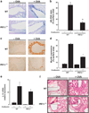
Ovalbumin (OVA)-induced mucin production is blunted in Ire1β−/− mice. (a) Alcian Blue–Periodic Acid Schiff (AB–PAS) staining of mucous cells in wild-type (WT) and Ire1β−/− mice exposed to saline (−OVA) or OVA. (b) Quantification of mucous cell expression as a percentage of AB–PAS stain/surface area of airway epithelia. Data were derived from large airways. n = 3 animals per group. *P < 0.05, Ire1β−/− vs. WT mice exposed to OVA. (c) Muc5b staining in airway epithelia from WT and Ire1β−/− mice exposed to saline (−OVA) or OVA. (d) Quantification of Muc5b expression in large airways as a percentage of Muc5b stain/surface area of airway epithelia. n = 3 animals per group. *P < 0.05, Ire1β−/− vs. WT mice exposed to OVA. (e,f) WT and Ire1β−/− mice exhibit a similar OVA-induced inflammatory response (e: OVA-upregulated interleukin (IL)-13 mRNA expression; (f) OVA-induced eosinophilic airway infiltration). (f) Insets illustrate the absence (left panels) or presence (right panels) of eosinophils infiltrating the airways.
Figure 6
Figure 6
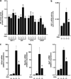
Endoplasmic reticulum (ER) stress and mucous cell gene expression in saline- and OVA-challenged wild-type (WT) vs. IRE1β−/− mice. (a) mRNA expression levels of total X-box binding protein-1 (XBP-1), immunoglobulin binding protein (BIP)/GRP78, activating transcription factor 4 (ATF4), and CHOP in tracheas from saline and OVA-challenged WT vs. Ire1β−/− mice. (b) OVA increases XBP-1 mRNA splicing in WT mice, and this response is blunted in Ire1β−/− mice. *P < 0.05, Ire1β−/− vs. WT mice exposed to OVA. (c) OVA induces upregulation of mucous cell genes in WT mice, and this response is blunted in Ire1β−/− mice. *P < 0.05, Ire1β−/− vs. WT mice exposed to OVA. KO: Ire1β−/− (knockout) mice.
Figure 7
Figure 7
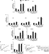
Inositol-requiring enzyme 1 (IRE1)β gene expression is associated with the expression of mucous cell genes in Calu-3 cell cultures. Time courses for 50 ng ml−1 serosal interleukin (IL)-13-modulated IRE1β and IRE1α gene expression (a), immunoglobulin binding protein (BIP)/GRP78, activating transcription factor 4 (ATF4) and CHOP, (b) and MUC5B and MUC5AC (c) in polarized cultures of Calu-3 cells stably expressing a control pSIREN vector (black bars) or a pSIREN vector containing a IRE1β shRNA (white bars). Data depict reverse transcriptase-PCR (RT-PCR) analyses. Data are from four experiments. *P < 0.05, pSIREN IRE1β shRNA-expressing cells vs. pSIREN control-expressing cells. (d; left): Correlation coefficient analysis between IRE1β gene expression and SAM pointed domain containing ETS transcription factor (SPDEF; P-value for correlation is shown on top of figure). (d; right): Time courses for 50 ng ml−1 serosal IL-13-modulated SPDEF gene expression in polarized cultures of Calu-3 cells stably expressing a control pSIREN vector (black bars) or a pSIREN vector containing a IRE1β shRNA (white bars). Data depict RT-PCR analyses. (e; left): Correlation coefficient analysis between IRE1β gene expression and anterior gradient homolog 2 (AGR2) gene expression (P-value for correlation is shown on top of figure). (e; right): Western blot (representative from three experiments) depicting the time course for 50 ng ml−1 serosal IL-13-induced AGR2 protein expression from whole-cell lysates from polarized cultures of Calu-3 cells stably expressing the control vector (“−” on top of figure) or IRE1β shRNA (“+” on top of figure). β-actin is used as a loading control.
Figure 8
Figure 8
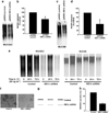
Reduction of inositol-requiring enzyme 1 (IRE1) β decreases basal and interleukin (IL)-13-stimulated cellular mucin content and mucin secretion. (a) Western blot from a 1% agarose gel depicting the staining for basal levels of MUC5AC protein expression from whole-cell lysates from polarized cultures of Calu-3 cell expressing a pSIREN control vector or a pSIREN vector containing a IRE1β shRNA. Data are representative of three individual experiments. (b) Compiled data from the MUC5AC signals from a. (c) Western blot from a 1% agarose gel depicting the staining for basal levels of MUC5B protein expression from whole-cell lysates from polarized Calu-3 cell cultures expressing a pSIREN control vector or a pSIREN vector containing an IRE1β shRNA. Data are representative of three individual experiments. (d) Compiled data from the MUC5B signals from c. (e) Western blots from 1% agarose gels depicting the time course for 50 ng ml−1 serosal IL-13-induced MUC5AC (left) and MUC5B (right) protein expression from whole-cell lysates from polarized Calu-3 cell cultures expressing a pSIREN control vector or a pSIREN vector containing an IRE1β shRNA. (f) Immunocytochemical detection of MUC5AC from Calu-3 cell cultures expressing a pSIREN control vector (left) or a pSIREN vector containing a IRE1β shRNA (right). Bar (right panel) = 20 µm. (g) Slot blots for basal MUC5AC secreted protein in Calu-3 cells expressing a pSIREN vector containing a IRE1β shRNA vs. Calu-3 cells expressing a pSIREN control vector. (h) Compiled data from the MUC5AC signals depicted in g. Data are representative of three to four experiments. *P < 0.05, pSIREN IRE1β shRNA-expressing cells vs. pSIREN control-expressing cells.
Figure 9
Figure 9
Inositol-requiring enzyme 1 (IRE1) α knockdown does not affect mucin production in Calu-3 cells. (a)mRNAlevels of IRE1α, IRE1β,MUC5AC, and MUC5B in Calu-3 airway epithelial cultures expressing a control vector (black bars) or a vector containing an IRE1α shRNA (white bars). (b) Immunocytochemical detection of MUC5AC from Calu-3 cell cultures expressing a control vector (left) or a vector containing an IRE1α shRNA (right). Bar (left panel) = 20 µm. (c) Slot blot for basal MUC5AC secreted protein in Calu-3 cells expressing a vector containing an IRE1β shRNA vs. Calu-3 cells expressing a control vector. (d) Compiled data from the MUC5AC signals depicted in c. Data are representative of three experiments.
Figure 10
Figure 10
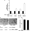
Overexpression of inositol-requiring enzyme 1 (IRE1) β increases cellular mucin content and basal mucin secretion. (a) IRE1β mRNA levels in Calu-3 cell cultures expressing a control pQCXIN vector or a pQCXIN vector containing IRE1β. (b) Immunocytochemical detection of MUC5AC from Calu-3 cell cultures expressing a control pQCXIN vector (left) or a pQCXIN vector containing IRE1β (right). Bar (left panel) = 20 µm. (c) Slot blots for basal MUC5AC secreted protein in Calu-3 cells expressing a pQCXIN vector containing IRE1β vs. Calu-3 cells expressing a control pQCXIN vector. (d) Compiled data from the MUC5AC signals depicted in c. *P < 0.05, Calu-3 cells expressing a pQCXIN vector containing IRE1β vs. Calu-3 cells expressing a control pQCXIN vector. Data are representative of three to four individual experiments.
Figure 11
Figure 11
Inositol-requiring enzyme 1 (IRE1) β-dependent mucin production is mediated by activation of X-box binding protein-1 (XBP-1) coupled to upregulation of anterior gradient homolog 2 (AGR2). (a) Representative Southern blot illustrating that interleukin (IL)-13 triggers XBP-1mRNA splicing in Calu-3 cell cultures expressing a pSIREN control vector, and this function is blunted in cultures expressing a pSIREN vector containing a IRE1β shRNA. (b) Compilation of the XBP-1 mRNA splicing data expressed as a percentage of XBP-1 mRNA splicing from t = 0 from control cultures. Data were derived from the same cDNA samples used in Figure 7 to assess the mRNA levels of IRE1β. (c): AGR2 mRNA expression in Calu-3 cells expressing a control pQCXIN vector, a pQCXIN vector containing spliced XBP-1, or a pQCXIN vector containing a dominant-negative XBP-1 (DN-XBP-1). *P < 0.05, spliced XBP-1-expressing cells vs. control cells; # P < 0.05, DN-XBP-1-expressing cells vs. control cells. (d) Immunocytochemical detection of MUC5AC from Calu-3 cell cultures expressing a control pQCXIN vector (left), a pQCXIN vector containing spliced XBP-1 (center), or a pQCXIN vector containing a DN-XBP-1 (right). Bar (left figure) = 20 µm. (e) Slot blots illustrating the basal levels of MUC5AC secreted protein in Calu-3 cells expressing a control pQCXIN vector, a pQCXIN vector containing spliced XBP-1, and a pQCXIN vector containing a DN-XBP-1. (f) Compiled data from the MUC5AC signals depicted in e. *P < 0.05, spliced XBP-1- or DN-XBP-1-expressing cells vs. control cells; # P < 0.05, spliced XBP-1- vs. DN-XBP-1-expressing cells. Data are from three experiments.

Similar articles

Cited by

References

    1. Kirkham S, Sheehan JK, Knight D, Richardson PS, Thornton DJ. Heterogeneity of airways mucus: variations in the amounts and glycoforms of the major oligomeric mucins MUC5AC and MUC5B. Biochem. J. 2002;361(Part 3):537–546. - PMC - PubMed
    1. Morcillo EJ, Cortijo J. Mucus and MUC in asthma. Curr. Opin. Pulm. Med. 2006;12:1–6. - PubMed
    1. Ribeiro CMP, Paradiso AM, Carew MA, Shears SB, Boucher RC. Cystic fibrosis airway epithelial Ca2 + i signaling. The mechanism for the larger agonist-mediated Ca2 + i signals in human cystic fibrosis airway epithelia. J. Biol. Chem. 2005;280:10202–10209. - PubMed
    1. Ribeiro CMP. Chronic airway infection/Inflammation Induces a Ca2+ i-dependent hyperinflammatory response in human cystic fibrosis airway epithelia. J. Biol. Chem. 2005;280:17798–17806. - PubMed
    1. Martino MEB, Olsen JC, Fulcher NB, Wolfgang MC, O’Neal WK, Ribeiro CMP. Airway epithelial inflammation-induced endoplasmic reticulum Ca(2+) store expansion ismediated by X-box binding protein-1. J. Biol. Chem. 2009;284:14904–14913. - PMC - PubMed

Publication types

MeSH terms

  NODES
INTERN 1
twitter 2