Right and left ventricular diastolic pressure-volume relations: a comprehensive review
- PMID: 23179133
- PMCID: PMC3594525
- DOI: 10.1007/s12265-012-9424-1
Right and left ventricular diastolic pressure-volume relations: a comprehensive review
Abstract
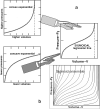
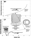
Ventricular compliance alterations can affect cardiac performance and adaptations. Moreover, diastolic mechanics are important in assessing both diastolic and systolic function, since any filling impairment can compromise systolic function. A sigmoidal passive filling pressure-volume relationship, developed using chronically instrumented, awake-animal disease models, is clinically adaptable to evaluating diastolic dynamics using subject-specific micromanometric and volumetric data from the entire filling period of any heartbeat(s). This innovative relationship is the global, integrated expression of chamber geometry, wall thickness, and passive myocardial wall properties. Chamber and myocardial compliance curves of both ventricles can be computed by the sigmoidal methodology over the entire filling period and plotted over appropriate filling pressure ranges. Important characteristics of the compliance curves can be examined and compared between the right and the left ventricle and for different physiological and pathological conditions. The sigmoidal paradigm is more accurate and, therefore, a better alternative to the conventional exponential pressure-volume approximation.
Figures


Similar articles
-
Evaluation of right and left ventricular diastolic filling.J Cardiovasc Transl Res. 2013 Aug;6(4):623-39. doi: 10.1007/s12265-013-9461-4. Epub 2013 Apr 13. J Cardiovasc Transl Res. 2013. PMID: 23585308 Free PMC article. Review.
-
Right ventricular diastolic function in canine models of pressure overload, volume overload, and ischemia.Am J Physiol Heart Circ Physiol. 2002 Nov;283(5):H2140-50. doi: 10.1152/ajpheart.00462.2002. Am J Physiol Heart Circ Physiol. 2002. PMID: 12384492 Free PMC article.
-
The effects of left heart assist on right ventricular muscle mechanics and ventricular coupling in the injured heart.J Thorac Cardiovasc Surg. 1994 Sep;108(3):477-86. J Thorac Cardiovasc Surg. 1994. PMID: 8078340
-
Effects of gradual volume loading on left ventricular diastolic function in dogs: implications for the optimisation of cardiac output.Heart. 1996 Apr;75(4):352-7. doi: 10.1136/hrt.75.4.352. Heart. 1996. PMID: 8705760 Free PMC article.
-
Assessment of left-ventricular function.Thorac Cardiovasc Surg. 1998 Sep;46 Suppl 2:248-54. doi: 10.1055/s-2007-1013081. Thorac Cardiovasc Surg. 1998. PMID: 9822175 Review.
Cited by
-
Challenges and Controversies in Hypertrophic Cardiomyopathy: Clinical, Genomic and Basic Science Perspectives.Rev Esp Cardiol (Engl Ed). 2018 Mar;71(3):132-138. doi: 10.1016/j.rec.2017.07.003. Epub 2017 Aug 10. Rev Esp Cardiol (Engl Ed). 2018. PMID: 28802532 Free PMC article.
-
Clinical-pathological correlations of BAV and the attendant thoracic aortopathies. Part 2: Pluridisciplinary perspective on their genetic and molecular origins.J Mol Cell Cardiol. 2019 Aug;133:233-246. doi: 10.1016/j.yjmcc.2019.05.022. Epub 2019 Jun 6. J Mol Cell Cardiol. 2019. PMID: 31175858 Free PMC article. Review.
-
Evaluation of right and left ventricular diastolic filling.J Cardiovasc Transl Res. 2013 Aug;6(4):623-39. doi: 10.1007/s12265-013-9461-4. Epub 2013 Apr 13. J Cardiovasc Transl Res. 2013. PMID: 23585308 Free PMC article. Review.
-
Genomic translational research: Paving the way to individualized cardiac functional analyses and personalized cardiology.Int J Cardiol. 2017 Mar 1;230:384-401. doi: 10.1016/j.ijcard.2016.12.097. Epub 2016 Dec 21. Int J Cardiol. 2017. PMID: 28057368 Free PMC article. Review.
-
Assessment of myocardial viscoelasticity with Brillouin spectroscopy in myocardial infarction and aortic stenosis models.Sci Rep. 2021 Nov 1;11(1):21369. doi: 10.1038/s41598-021-00661-4. Sci Rep. 2021. PMID: 34725389 Free PMC article.
References
-
- Zhang SJ, Truskey GA, Kraus WE. Effect of cyclic stretch on1D-integrin expression and activation of FAK and RhoA. Am J Physiol Cell Physiol. 2007;292:C2057–C2069. - PubMed
-
- Pasipoularides A. Heart's vortex: intracardiac blood flow phenomena. People's Medical Publishing House; Shelton, CT: 2010. p. 960.
-
- Mirsky I, Pasipoularides A. Clinical assessment of diastolic function. Prog Cardiovasc Dis. 1990;32:291–318. - PubMed
-
- Hayley BD, Burwash IG. Heart failure with normal left ventricular ejection fraction: role of echocardiography. Cur Opin Cardiol. 2012;27:169–180. - PubMed
Publication types
MeSH terms
Grants and funding
LinkOut - more resources
Full Text Sources

