. 2012;7(12):e49789.
doi: 10.1371/journal.pone.0049789. Epub 2012 Dec 14.

Dimerization with cannabinoid receptors allosterically modulates delta opioid receptor activity during neuropathic pain

Affiliations

Dimerization with cannabinoid receptors allosterically modulates delta opioid receptor activity during neuropathic pain

Ittai Bushlin et al. PLoS One. 2012.

Abstract

The diversity of receptor signaling is increased by receptor heteromerization leading to dynamic regulation of receptor function. While a number of studies have demonstrated that family A G-protein-coupled receptors are capable of forming heteromers in vitro, the role of these heteromers in normal physiology and disease has been poorly explored. In this study, direct interactions between CB(1) cannabinoid and delta opioid receptors in the brain were examined. Additionally, regulation of heteromer levels and signaling in a rodent model of neuropathic pain was explored. First we examined changes in the expression, function and interaction of these receptors in the cerebral cortex of rats with a peripheral nerve lesion that resulted in neuropathic pain. We found that, following the peripheral nerve lesion, the expression of both cannabinoid type 1 receptor (CB(1)R) and the delta opioid receptor (DOR) are increased in select brain regions. Concomitantly, an increase in CB(1)R activity and decrease in DOR activity was observed. We hypothesize that this decrease in DOR activity could be due to heteromeric interactions between these two receptors. Using a CB(1)R-DOR heteromer-specific antibody, we found increased levels of CB(1)R-DOR heteromer protein in the cortex of neuropathic animals. We subsequently examined the functionality of these heteromers by testing whether low, non-signaling doses of CB(1)R ligands influenced DOR signaling in the cortex. We found that, in cortical membranes from animals that experienced neuropathic pain, non-signaling doses of CB(1)R ligands significantly enhanced DOR activity. Moreover, this activity is selectively blocked by a heteromer-specific antibody. Together, these results demonstrate an important role for CB(1)R-DOR heteromers in altered cortical function of DOR during neuropathic pain. Moreover, they suggest the possibility that a novel heteromer-directed therapeutic strategy for enhancing DOR activity, could potentially be employed to reduce anxiety associated with chronic pain.


Conflict of interest statement

Competing Interests: The authors have declared that no competing interests exist.

Figures

Figure 1
Figure 1. Lesioned animals experience mechanical allodynia.
Mechanical response threshold to Von Frey fibers was measured before and after surgery in sham and L5SNT lesioned animals. Response threshold value was calculated as described in “Methods”. Data represent Mean ± SEM (n = 12 animals/group).
Figure 2
Figure 2. CB1R levels increase in cortex of lesioned animals.
A, Representative Western Blot and quantification for CB1R and CNX (Calnexin) in cortical membranes from 3 individual sham or lesioned animals, 14 days after surgery. Data represent Mean ± SEM (n = 6 animals/group). B, ELISA data for CB1R detection using a CB1R specific antibody in cortical membranes from sham and lesioned animals 14 days after surgery. Data represent Mean ± SEM (n = 4 animals/group). Statistically significant differences between sham and lesion groups are indicated*, p<0.05; **, p<0.01 (t-test). C, RT-PCR for CB1R (measured relative to GAPDH) in cortical preparations from sham and lesioned animals 14 days after surgery. Data represent Mean ± SEM (n = 5 animals/group). Statistically significant differences between sham and lesion groups are indicated *, p<0.05 (t-test).
Figure 3
Figure 3. CB1R levels are increased in cortex of lesioned animals as visualized by immunohistochemistry.
A,B A representative pair of images from studies examining the localization of CB1R by immunohistochemistry in layer II/III of PFC of sham and lesioned animals 14 days after surgery. Representative of 8 images taken per condition is shown. Scale bar = 20 µm. Increased CB1R immunoreactivity is indicated by short arrows and cell bodies are indicated by the long arrow. C, Quantification of changes in CB1R fluorescence intensity in PFC. Data represents mean fluorescence intensity from 16 images (4 per animal, n = 2 animals/group; p<0.05, t-test).
Figure 4
Figure 4. DOR levels increase in cortex of lesioned animals.
A, Representative Western Blot and quantification for DOR and CNX in cortical membranes from 3 individual sham or lesioned animals. Data represent Mean ± SEM (n = 6 animals/group). Statistically significant differences between sham and lesion groups are indicated **, p<0.01 (t-test). B, ELISA data for DOR in cortical membranes from sham and lesioned animals. Data represent Mean ± SEM (n = 4 animals/group). Statistically significant differences between sham and lesion groups are indicated ***, p<0.001 (t-test).
Figure 5
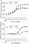
Figure 5. CB1R activity increases while DOR activity decreases in cortex of lesioned animals.
A, [35S]GTPγS binding was carried out with cortical membranes from sham and lesioned animals. Membranes from cortices were prepared as described in “Methods” and treated with 0.1 pM – 10 µM Hu-210 for 1 hour. [35S]GTPγS binding to membranes was detected using a scintillation counter. Basal [35S]GTPγS binding in vehicle treated membranes is taken as 100%. Data represent Mean ± SEM (n = 3 individual animals in triplicate). B, Membranes from cortices of sham and lesioned animals were treated with 1 pM – 10 µM DPDPE for 1.5 hours. [35S]GTPγS binding to membranes was detected using a scintillation counter. Basal [35S]GTPγS binding in vehicle treated membranes is taken as 100%. Data represent Mean ± SEM (n = 3 individual animals in triplicate).
Figure 6
Figure 6. CB1R-DOR antibody specificity and increases in CB1R-DOR levels during neuropathic pain.
A, ELISA for mouse monoclonal CB1R-DOR antibody using the following cell lines: 1) HEK cells expressing the following receptor combinations: DOR, KOR+DOR, or MOR+DOR; 2) N2A cells (which endogenously express CB1R) alone or co-expressing the following receptors: AT1R, CB2R, DOR, MOR, or KOR. Data represent Mean ± SEM (n = 3 independent experiments in triplicate). Statistically significant differences between sham and lesion groups are indicated ***, p<0.001 (t-test). B, ELISA for rat polyclonal DOR, and mouse monoclonal CB1R-DOR antibodies in cortical membranes from wild type, CB1R −/−, and DOR −/− animals. Data represent Mean ± SEM (n = 3 animals/group). Statistically significant differences between wild type and −/− groups are indicated ***, p<0.001 (t-test). C, ELISA for CB1R-DOR in cortical membranes from sham and lesioned animals. Data represent Mean ± SEM (n = 4 animals/group). Statistically significant differences between sham and lesion groups are indicated ***, p<0.001 (t-test).
Figure 7
Figure 7. DOR activity is enhanced in the presence of CB1R ligands in cortical membranes from lesioned animals.
A, Membranes from cortices of lesioned animals were treated with 10 pM – 10 µM DPDPE in the absence of presence of 1 pM Hu-210, or with 1 pM Hu-210 alone for 1.5 hours. [35S]GTPγS binding to membranes was detected using a scintillation counter. Basal [35S]GTPγS binding in vehicle treated membranes is taken as 100%. Data represent Mean ± SEM (n = 3 individual animals in triplicate). Statistically significant differences between 10 µM DPDPE alone and 10 µM DPDPE+1 pM Hu-210 are indicated ***, p<0.001, (t test). B, Membranes from cortices of sham and lesioned animals were treated with 10 µM DPDPE in the absence of presence of 1 pM Hu-210, or with 1 pM Hu-210 alone for 1.5 hours. [35S]GTPγS binding to membranes was detected using a scintillation counter. Basal [35S]GTPγS binding in vehicle treated membranes is taken as 100%. Data represent Mean ± SEM (n = 4 individual animals in triplicate). Statistically significant differences between 10 µM DPDPE alone and 10 µM DPDPE+1 pM Hu-210 are indicated **, p<0.01, (t test). C, Membranes from cortices of lesioned animals were treated with 10 µM DPDPE in the absence of presence of 1 µM PF-514273, or with 1 µM PF-514273 alone for 1.5 hours. [35S]GTPγS binding to membranes was detected using a scintillation counter. Basal [35S]GTPγS binding in vehicle treated membranes is taken as 100%. Data represent Mean ± SEM (n = 3 individual animals in triplicate). Statistically significant differences between 10 µM DPDPE alone and 10 µM DPDPE+1 µM PF-514273 are indicated *, p<0.05, (t test). D, Membranes from cortices of lesioned animals were treated with 10 µM DPDPE in the absence of presence of 1 pM Hu-210, or 10 nM DAMGO or 10 nM U69593 for 1.5 hours. [35S]GTPγS binding to membranes was detected using a scintillation counter. Basal [35S]GTPγS binding in vehicle treated membranes is taken as 100%. Data represent Mean ± SEM (n = 3 individual animals in triplicate). Statistically significant differences between 10 µM DPDPE and 10 µM DPDPE+ligand are indicated **, p<0.01, (t test).
Figure 8
Figure 8. CB1R-DOR heteromer-specific antibody blocks enhancement of DOR activity.
A, Membranes from cortices of lesioned animals were treated with 10 µM DPDPE without or with Hu-210 in the absence or presence of 1 µg of indicated antibodies. [35S]GTPγS binding to membranes was detected using a scintillation counter. Basal [35S]GTPγS binding in vehicle treated membranes is taken as 100%. Data represent Mean ± SEM (n = 3 individual animals in triplicate). Statistically significant differences between 10 µM DPDPE+1 pM Hu-210 and 10 µM DPDPE+1 pM Hu-210+1 µg antibody are indicated ***, p<0.001, (t test). B, In the absence or presence of 1 µg CB1R-DOR heteromer-specific antibody, membranes from cortices of lesioned animals were treated with 10 µM DPDPE without or with 1 pM Hu-210. [35S]GTPγS binding to membranes was detected using a scintillation counter. Basal [35S]GTPγS binding in vehicle treated membranes is taken as 100%. Data represent Mean ± SEM (n = 3 individual animals in triplicate). Statistically significant differences between 10 µM DPDPE and 10 µM DPDPE+1 pM Hu-210 are indicated ***, p<0.001, (t test). C, Membranes from hippocampi of lesioned animals were treated with 10 µM DPDPE in the absence of presence of 1 pM Hu-210 for 1.5 hours. Membranes were also treated with 10 µM DPDPE and 1 pM Hu-210 in the presence of 1 µg of CB1R-DOR mAb. [35S]GTPγS binding to membranes was detected using a scintillation counter. Basal [35S]GTPγS binding in vehicle treated membranes is taken as 100%. Data represent Mean ± SEM (n = 3 individual animals in triplicate). n.s. not significant.
Figure 9
Figure 9. DOR binding is enhanced in the presence of low dose CB1R ligand in cortical membranes.
Membranes from cortices of sham and lesioned animals were treated with 0.5 nM [3H]DPDPE in the presence or absence of 1 pM Hu-210 for 1 hour. Ligand binding assay was carried out as described in “Methods” and [3H]DPDPE binding to membranes was detected using a scintillation counter. Data represent Mean ± SEM (n = 7 animals/group). Statistically significant differences between [3H]DPDPE alone and [3H]DPDPE+1 pM Hu-210 are indicated ***, p<0.001, (t test).
Figure 10
Figure 10. DOR binding is enhanced by CB1R ligands in membranes of cells expressing CB1R and DOR.
A, Membranes from N2A-DOR cells were incubated with 100 fM – 10 µM Hu-210 in the presence of 0.5 nM [3H]DPDPE ± 1 µg CB1R-DOR monoclonal antibody for 1 hour. Ligand binding assay was carried out as described in “Methods” and [3H]DPDPE binding to membranes was detected using a scintillation counter. Data represent Mean ± SEM (n = 3 experiments in triplicate). B, Membranes from N2A-DOR cells in which CB1R expression was decreased by siRNA transfection were treated with 100 fM – 10 µM Hu-210 in the presence of 0.5 nM [3H]DPDPE. Ligand binding assay was carried out as described in “Methods” and [3H]DPDPE binding to membranes was detected using a scintillation counter. Data represent Mean ± SEM (n = 3 experiments in triplicate). C, Membranes from N2A-DOR cells were treated with 100 fM – 10 µM PF-514273 in the presence of 0.5 nM [3H]DPDPE along with the presence or absence of 1 µg CB1R-DOR monoclonal antibody for 1 hour. Ligand binding assay was carried out as described in “Methods” and [3H]DPDPE binding to membranes was detected using a scintillation counter. Data represent Mean ± SEM (n = 3 experiments in triplicate).

Similar articles

Cited by

References

    1. Finnerup NB, Sindrup SH, Jensen TS (2010) The evidence for pharmacological treatment of neuropathic pain. Pain 150: 573–581. - PubMed
    1. O'Connor AB, Dworkin RH (2009) Treatment of neuropathic pain: an overview of recent guidelines. Am J Med 122: S22–32. - PubMed
    1. Bausch SB, Patterson TA, Appleyard SM, Chavkin C (1995) Immunocytochemical localization of delta opioid receptors in mouse brain. J Chem Neuroanat 8: 175–189. - PubMed
    1. Hohmann AG, Briley EM, Herkenham M (1999) Pre- and postsynaptic distribution of cannabinoid and mu opioid receptors in rat spinal cord. Brain Res 822: 17–25. - PubMed
    1. Maldonado R, Valverde O (2003) Participation of the opioid system in cannabinoid-induced antinociception and emotional-like responses. Eur Neuropsychopharmacol 13: 401–410. - PubMed

Publication types

MeSH terms

  NODES
twitter 2