. 2013 Mar;9(3):361-73.
doi: 10.4161/auto.23066. Epub 2013 Jan 4.

Regulation of nutrient-sensitive autophagy by uncoordinated 51-like kinases 1 and 2

Affiliations

Regulation of nutrient-sensitive autophagy by uncoordinated 51-like kinases 1 and 2

Fiona McAlpine et al. Autophagy. 2013 Mar.

Abstract

Macroautophagy, commonly referred to as autophagy, is a protein degradation pathway that occurs constitutively in cells, but can also be induced by stressors such as nutrient starvation or protein aggregation. Autophagy has been implicated in multiple disease mechanisms including neurodegeneration and cancer, with both tumor suppressive and oncogenic roles. Uncoordinated 51-like kinase 1 (ULK1) is a critical autophagy protein near the apex of the hierarchal regulatory pathway that receives signals from the master nutrient sensors MTOR and AMP-activated protein kinase (AMPK). In mammals, ULK1 has a close homolog, ULK2, although their functional distinctions have been unclear. Here, we show that ULK1 and ULK2 both function to support autophagy activation following nutrient starvation. Increased autophagy following amino acid or glucose starvation was disrupted only upon combined loss of ULK1 and ULK2 in mouse embryonic fibroblasts. Generation of PtdIns3P and recruitment of WIPI2 or ZFYVE1/DFCP1 to the phagophore following amino acid starvation was blocked by combined Ulk1/2 double knockout. Autophagy activation following glucose starvation did not involve recruitment of either WIPI1 or WIPI2 to forming autophagosomes. Consistent with a PtdIns3P-independent mechanism, glucose-dependent autophagy was resistant to wortmannin. Our findings support functional redundancy between ULK1 and ULK2 for nutrient-dependent activation of autophagy and furthermore highlight the differential pathways that respond to amino acid and glucose deprivation.

Keywords: MEF; ULK1; ULK2; WIPI1; WIPI2; knockout; nutrient starvation.


Figures
None
Figure 1. Loss of ULK1 and ULK2 inhibits autophagy following amino acid starvation. (A) Wild-type (WT), Ulk1 knockout (ULK1KO), Ulk2 knockout (ULK2KO) and two independently derived Ulk1/Ulk2 double knockout (DKO) MEF lines were treated with full-nutrient medium (F), EBSS (-AA) or EBSS containing 50 nM bafilomycin A1 (Baf) for 2 h. Resolved cell lysates were immunoblotted with antibodies against ULK1, actin and LC3 (Nanotools 5F10 monoclonal). “*” indicates nonspecific band on ULK1 immunoblot. MEF lines were immortalized with SV40 T antigen. Below: LC3-II/LC3-I signals were plotted as average ± SEM, (n = 4) using 2 different y-axis scales. Two-tailed paired t-tests were performed relative to values of WT MEFs under the same condition: *p < 0.04; **p < 0.005; ap = 0.14; bp = 0.056; cp = 0.12. (B) Total RNA was extracted from MEF lines described in (A) and analyzed for Ulk1 and Ulk2 transcript levels using quantitative RT-PCR. Plots show Ulk1 or Ulk2 levels normalized to Actin transcript levels (average ± SEM, (n = 3), values relative to WT cells). (C) WT and DKO MEF lines were treated as in (A) to full-nutrient medium or EBSS for 2 h. Resolved cell lysates were immunoblotted with antibodies against phospho-(Ser79) acetyl CoA carboxylase (P-ACC), phospho-(Ser240/244) ribosomal protein S6 (P-RPS6); actin and LC3 (5F10 monoclonal antibody). (D) MEF lines were treated to full-nutrient medium (F) or glucose starvation (−Glc) for 16 h before lysis and immunoblot analysis as in (C). (E) MEF lines were steady-state labeled overnight with C14-valine, chased for 24 h, and analyzed for protein degradation over 2 h of amino acid starvation. Plot shows average ± SEM (n = 3). Two-tailed paired t-tests were performed relative to WT MEFs: *p < 0.02; **p < 0.01. (F) WT and Ulk DKO#1 MEFs were treated as in (A) for 2 h and fixed for LC3 immunostaining. Shown are representative confocal images of −AA and −AA+Baf conditions. Scale bar: 10 μm. Plots show average puncta/cell ± SEM (each column: n = 22 to 39 cells per condition from two experiments). Two-tailed paired t-tests when compared with WT MEFs under the same condition: ***p < 0.0003; *p < 0.02. (G) Primary WT and DKO MEFs were subjected to starvation conditions as in (A) and lysed for immunoblot analysis of SQSTM1 and actin. Right: Plotted densitometry [SQSTM1 levels normalized to actin, shown as average ± SEM (n = 4)]. Two-tailed paired t-tests when compared with WT MEFs under same conditions: *p < 0.04, **p < 0.01.
None
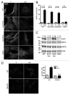
Figure 2. ULK1 and ULK2 are both required for WIPI2 spot formation following amino acid starvation. (A) WT, ULK1KO, ULK2KO and DKO primary MEFs were treated with either full nutrient-medium (F) or EBSS (−AA) for 2 h before fixation. Cells were then stained with an antibody against WIPI2. (B) In 10 fields per condition (two independent experiments) Hoechst-stained nuclei (cell number) and WIPI2 puncta were quantified (WIPI2 puncta/cell ± SEM) (n = 20 fields). Differences between F and −AA are indicated by ****p < 0.0001 by one-way ANOVA with Bonferroni post hoc test, n.s. non-significant. (C) Primary MEF lines were treated as in (A). Cell lysates were immunoblotted for ULK1, WIPI1, WIPI2 and actin. (D) WT and DKO#1 immortalized cells stably expressing GFP-ZFYVE1 were treated to full-nutrient medium (F) or EBSS (−AA) for 2 h before fixation. (A and D) Scale bars: 10 μm. Plot shows puncta/cell (average ± SEM for n = 29 to 37 cells per condition from two experiments). Two-tailed paired t-test comparing to WT MEFs under same conditions: ***p < 0.0001.
None
Figure 3. ATG13 knockdown phenocopies Ulk1/Ulk2 double knockout for the LC3 lipidation pathway following amino acid starvation. (A) WT and DKO primary MEFs were transfected with either RISC-Free control or mouse Atg13-_targeting siRNA. Following knockdown, cells were treated with either full-nutrient medium (F), EBSS (−AA) or EBSS containing bafilomycin A1 (−AA+Baf) (100 nM) for 2 h. Resolved cell lysates were immunoblotted with antibodies against ULK1, ATG13, actin and LC3 (Abcam polyclonal). (B) Average LC3-II/actin densitometry ± SEM (n = 4). Two-tailed paired t-tests were performed relative to WT-RISC-Free siRNA MEFs under the same conditions: ap = 0.067; bp = 0.068; cp = 0.089.
None
Figure 4. ATG13 knockdown phenocopies Ulk1/Ulk2 double knockout on the formation of WIPI2-positive autophagosomal membranes following amino acid starvation. WT, ULK1KO, ULK2KO and DKO primary MEFs were transfected with either RISC-Free control or mouse Atg13-_targeting siRNA. Following knockdown, cells were treated with either full-nutrient medium (F) or EBSS (−AA) for 2 h before fixation. Cells were stained with an antibody against WIPI2. Scale bar:10 μm.
None
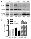
Figure 5. Combined Ulk1 and Ulk2 knockout reduces the LC3 lipidation response to glucose starvation. (A) WT or DKO primary MEFs were treated to either full-nutrient medium (F), glucose-free conditions (−Glc), or glucose-free medium + bafilomycin A1 (−Glc+Baf) (100 nM) for 24 h. Resolved cell lysates were immunoblotted with antibodies against ULK1, actin and LC3 (Abcam polyclonal). “*” indicates nonspecific band. (B) LC3-II and actin signals were quantified by densitometry. Results are shown as average ± SEM (n = 4). Two-tailed paired t-tests were performed relative to WT MEFs under the same treatment: *p < 0.02.
None
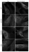
Figure 6. Glucose starvation does not stimulate production of WIPI2-containing membranes. WT, ULK1KO, ULK2KO and DKO primary MEFs were treated with either full-nutrient medium (Fed) or glucose-free medium (glucose starved) for 24 h then fixed. Cells were stained with an antibody against WIPI2. Scale bar:10 μm.
None
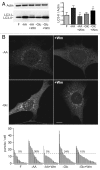
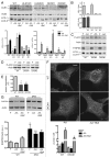
Figure 7. LC3 lipidation and autophagosome formation following glucose starvation are PtdIns3P-independent. (A) WT primary MEFs were treated with full-nutrient medium (F), EBSS (−AA), or EBSS containing 100 nM wortmannin (−AA+Wm) for 2 h. Alternatively, cells were incubated in glucose-free medium (−Glc) or glucose-free medium containing 100 nM wortmannin (−Glc+Wm) for 24 h. Resolved cells lysates were immunoblotted with antibodies for LC3 (Abcam polyclonal) and actin. Results are shown as average LC3-II/actin ± SEM (n = 4). Two-tailed paired t-tests were performed relative to equivalent treatment condition without wortmannin: *p < 0.04. (B) WT (immortalized) MEFs were treated with full-nutrient medium (F) or EBSS (−AA) for 2 h. Alternatively, cells were incubated in glucose-free medium (−Glc) for 16 h. Where indicated, incubations included 100 nM wortmannin (+Wm). Cells were fixed after treatments for LC3 immunostaining. Shown are representative confocal images of −AA and −Glc conditions ± wortmannin. Scale bar: 10 μm. Plot shows puncta/cell distributions for each condition. Each column represents quantification of one cell (40–60 cells/condition taken from two experiments). Shown are percentages of cells within each group containing 15 (or more) puncta/cell.
None
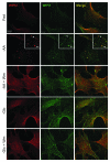
Figure 8. Absence of WIPI1- and WIPI2-positive autophagosomal membranes following glucose starvation. WT primary MEFs were incubated in full-nutrient medium (Fed), EBSS (−AA), or EBSS with 100 nM wortmannin (−AA+Wm) for 2 h. Alternatively, cells were glucose starved without or with 100 nM wortmannin (−Glc, −Glc+Wm) for 24 h before fixation. Cells were stained with antibodies against WIPI2 (red) and WIPI1 (green). Scale bar: 10 μm. Zoomed inset shows WIPI2 and WIPI1 colocalization following amino acid starvation (arrow: strong colocalization; arrowhead: partial).

Similar articles

Cited by

References

    1. Tooze SA, Jefferies HB, Kalie E, Longatti A, McAlpine FE, McKnight NC, et al. Trafficking and signaling in mammalian autophagy. IUBMB Life. 2010;62:503–8. doi: 10.1002/iub.334. - DOI - PubMed
    1. Orsi A, Polson HE, Tooze SA. Membrane trafficking events that partake in autophagy. Curr Opin Cell Biol. 2010;22:150–6. doi: 10.1016/j.ceb.2009.11.013. - DOI - PubMed
    1. Mizushima N, Yoshimori T, Ohsumi Y. The role of Atg proteins in autophagosome formation. Annu Rev Cell Dev Biol. 2011;27:107–32. doi: 10.1146/annurev-cellbio-092910-154005. - DOI - PubMed
    1. Yan J, Kuroyanagi H, Kuroiwa A, Matsuda Y, Tokumitsu H, Tomoda T, et al. Identification of mouse ULK1, a novel protein kinase structurally related to C. elegans UNC-51. Biochem Biophys Res Commun. 1998;246:222–7. doi: 10.1006/bbrc.1998.8546. - DOI - PubMed
    1. Kuroyanagi H, Yan J, Seki N, Yamanouchi Y, Suzuki Y, Takano T, et al. Human ULK1, a novel serine/threonine kinase related to UNC-51 kinase of Caenorhabditis elegans: cDNA cloning, expression, and chromosomal assignment. Genomics. 1998;51:76–85. doi: 10.1006/geno.1998.5340. - DOI - PubMed

Publication types

MeSH terms

  NODES
twitter 2