. 2012;1(3):316-33.
Epub 2012 Nov 25.

Multiple mechanisms of extracellular tau spreading in a non-transgenic tauopathy model

Affiliations

Multiple mechanisms of extracellular tau spreading in a non-transgenic tauopathy model

Meghan N Le et al. Am J Neurodegener Dis. 2012.

Abstract

While the interneuronal propagation of neurofibrillary lesions in Alzheimer's disease and other tauopathies now appears to involve the spreading of tau-associated toxicity, little is known about its mechanism. We characterized the movement of human tau through the brain of a non-transgenic lower vertebrate tauopathy model in which full-length wild type and mutant human tau isoforms were expressed in identified neurons, thus permitting the identification and localization of EC tau sources. We describe two distinct patterns of tau spreading that correspond to tau species that lack (MTBR-) and contain (MTBR+) the tau microtubule-binding region. These patterns illustrate the production, migration and uptake of EC tau and resemble some of the extracellular tau deposits typically seen in human brain after repeated traumatic injury in cases of chronic traumatic encephalopathy (CTE). We propose that misprocessed human tau can spread between CNS neurons via a variety of non-synaptic mechanisms as well as synaptically mediated mechanisms.

Keywords: CSF-tau; chronic traumatic encephalopathy; interneuronal lesion spread; neuron death; tau secretion.


Figures

Figure 1
Figure 1
Cellular and extracellular pathways by which human tau may be misprocessed in tauopathy models and in human neurodegenerative disease. In the mature CNS, tau is primarily associated with MTs in neuronal axons, where it is not toxic or associated with neurofibrillary pathology. Conditions predisposing to tau misprocessing include the generation of toxic levels of the Aβ peptide in AD, the presence of exonic point mutations that cause familial tauopathies, elevated baseline tau expression associated with the H1 haplotype, and repetitive brain trauma, which leads to tauopathy (CTE) and is associated with elevated AD risk. All of these can result in increased levels of non-MT associated tau, which may then be mislocated and misprocessed, leading to tau toxicity, stoichiometrically high levels of phosphorylation, altered tertiary structure and susceptibility to proteolytic cleavage by calpains and caspases. Misprocessing and toxicity are associated with the generation and appearance in th CSF of both N terminal tau fragments that lack the microtubule binding region of tau (MTBR- tau, red) or full length of C terminally cleaved tau species (MTBR+ tau, blue) that possess the MTBR. Interneuronal spreading of tau lesions may be due to direct transfer of toxic tau species between neurons followed by either a prionlike templated misfolding of tau in the receiving neuron or by Ca++ dysregulatory effects of oligomeric (MTBR+) tau or toxic N terminal (MTBR-) tau species. While tau lesion spreading is generally thought to occur in association with synapses, we propose that the periventricular and diffuse EC tau migration patterns described here illustrate additional pathways by which lesion spreading might occur in AD and in other tauopathies, including trauma –associated tauopathies such as CTE (arrows).
Figure 2
Figure 2
Distribution and quantitative analysis of diffuse and periventricular secreted tau patterns in the lamprey CNS. Panel A: Full length and N terminal half tau constructs were expressed cell autonomously via plasmid injection in lamprey ABCs for 5-40 days. All isoforms used lacked the E2 and E3 inserts. Full length isoforms contained 4 microtubule binding repeat motifs that either lacked (wild type) or contained a tauopathy-inducing point mutation within the second MTBR motif at position 301 (P301L) according to the numbering scheme for the longest of the 6 tau isoforms expressed in the human CNS. The N terminal construct used in this study (bottom) encodes residues 1-44 and 104-255 of human tau. The epitope site and phosphorylation sensitivity of antibodies used in this study. Phospho-specific sites are shown in red (serine/threonine) or green (tyrosine), with a dephosphorylation specific mAb (tau1) shown in blue. Phospho-independent Abs are shown in black. Panel B: Schematics summarizing the characteristic distribution of diffuse (top), focal (center) and periventricular (bottom) tau with respect to the ABC of origin (red, shown in cutaway images in the center of each panel). C: Live image of a lamprey brain (dorsal view) showing the expression of GFP-tagged tau in ABCs. The surface of the hindbrain is dominated by the central sulcus (CS) and the Sulci of His (SoH). Left center: Scoring sheet used in this study illustrating the semiquantitative analysis of the distribution of diffuse (red) and periventricular (PV) tau deposits in serially sectioned lamprey brains. Locations of cell bodies are shown in black. The distribution of PV tau was estimated as occupying a proportion of the ventricular surface for each section using the central sulcus and Sulcus of His as landmarks as shown. Anatomical landmarks such as the CS and SoH were frequently “skipped” (white arrow), suggesting that at least some periventricular tau transport occurs within the ventricle itself.
Figure 3
Figure 3
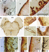
Examples of diffuse, focal and periventricular (PV) extracellular tau deposits in the lamprey brain. The relationship the deposits and their sites of origin from tau-expressing ABCs in the lamprey hindbrain are shown at left as indicated. Photomicrographs of examples of each EC deposit type from wild type (WT) and P301L tauopathy mutant (MTBR+) and a deletion construct (1-255) in which the MTBR region is not encoded (MTBR-) are shown. Scale bars: Diffuse, 100 μm; Focal and PV, 20 μm.
Figure 4
Figure 4
Quantitative analysis of EC tau gradients produced by ABCs expressing MTBR+ and MTBR- tau constructs.Diffuse, focal and periventricular tau deposits differ characteristically from each other in the degree to which they a)vary with respect to the construct expressed and b) with their site of origin in the expressing ABC (i.e. somatic, dendriticaxonal). Panel A: Periventricular (PV), focal and diffuse EC tau from each isoform used, immunostained with themAb Tau12, are shown. Panel B: The typical features of the tau12 immunolabel gradient for each type of EC tau isillustrated using a LUT that corresponds to ranges of DAB immunolabel intensity as shown at left. The images shownare derived from their corresponding image in Panel A. Note that while both diffuse and PV tau species are often intenselyimmunolabeled at their site of generation (shown as red in the LUT), diffuse deposits are spread over a muchlarger region of neuropil relative to their intensity that PV and focal deposits, suggesting that the former are significantlymore mobile. Panel C: Quantitative comparisons of gradient slopes (i.e. degree of intensity change per unitdistance from their site of origin - as defined in Methods) from each EC tau type are shown. Since the data distributionsexhibited noticeable skewness with respect to the mean value, both the Student’s t-test and the Fisher’s Exacttest were used to determine significance levels and gave similar results. Bars show the S.E.M. Left: All diffuse depositsexhibited significantly milder slopes than either PV or focal tau deposits, whereas focal deposits had significantlysteeper distribution slopes than “re-entering” PV gradients. Both somatic and axonal PV deposits were used in thisanalysis. Center Left: The absence of the MTBR from the construct expressed abolished focal and PV deposits,whereas its inactivation by pseudophosphorylation (S262D/S356D) made the slopes of fopcal deposits significantlyshallower. The presence of the P301L tauopathy mutation did not affect the distribution slope of any of the 3 EC taudeposit types. Center Right: Both focal and PV EC tau derived from ABC axons showed significantly steeper distributiongradients than did tau released from ABC somata. We saw no sign of low slope gradient generation (i.e. axonaldiffuse deposits) in this study. Right: While the overall extent of some EC deposits changed between 10 and 20-40days days of expression (see Figure 5), the slopes of all 3 EC deposit types were not significantly affected. Scale Barsare 100 μm, except for the inset high magnification at right, which is 20 μm.
Figure 5
Figure 5
PV and diffuse tau migration in the lamprey brain from the ABC of origin is modulated by the presence of the MTBR and/or the P301L point mutation. Panel A: Schematic representation of PV tau migration in the lamprey hindbrain. PV deposits typically originated from either the dorsal somata (top) and axons (bottom) of tau-expressing ABCs, and spread along the surface of the brain from the point of origin down a concentration gradient (illustrated by red, yellow and green zones, arrows). The numbers at right in both schematics show the slide numbers and the approximate sites of each of the individual sections shown in B. In some cases, maximal extents of PV tau were reached within the first 10 days of expression, with CE tau being present throughout the hindbrain at distances of at least 2 μm from the source somata (not shown). Panel B: Transverse sections taken from the sites indicated in A are shown and a semiquantitative representation of immunolabel intensity is shown for each section (right, see LUT at bottom). Somata (top) and axons (bottom) were identified as sources of EC tau by a) the observation of a focal gradient emerging from the dorsal soma or axon and b) the absence of other candidate sites in that section (or in adjacent sections). Scale bars: top: 50 μm; bottom, 200 μm. Panel C: Quantitative analysis of the “footprint” immunolabel maps for diffuse (top) and PV (bottom) tau. The incidence and extent of diffuse and PV tau within the lamprey brain were modulated by a) the presence of the MTBR, b) the P301L tauopathy mutation and c) the length of time over which tau was expressed in ABCs. We found that the absence of the MTBR resulted in exclusively diffuse secretion. This was present from the earliest time (5 days post plasmid injection) examined and thus confirmed the earlier study of Kim et. al (2010). By contrast, tau secretion was relatively restricted in lampreys expressing WT full length tau constructs, especially with respect to diffuse tau, which was secreted at low levels in a large minority of ABCs examined at all time-points. There was no significant increase in either the incidence or extent of diffuse EC tau with increasing time post plasmid injection in these animals (top, blue bars). The presence of the P301L point mutation (pink bars) greatly increased the extent of both diffuse and PV deposits. This was especially marked with diffuse deposits, which also became significantly more extensive over time. PV deposit extent also increased with time post injection, but this increase was not significant. Spreading of axonally secreted PV deposits (black bars) was not noticeably different from that of somatic origin, despite the difference in their slops (Figure 4C). Scale Bars: B (top) 50 μm, B (bottom) 200 μm.
Figure 6
Figure 6
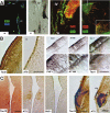
Phosphorylation state of EC tau species in the lamprey. PV tau was typically phosphorylated to a varyingextent at one or more of the classic “AD” sites in and around the MTBR, but many of the sections examined showedno significant immunostaining for phosphorylated tau (Ptau), especially at extracellular loci within the brain. Panel A:Confocal imaging of sections adjacent to Tau12/DAB immunolabeled sections shows some MTBR+ character andsome phosphorylation of all EC tau species, but especially PV (Panels A, left, Panel B), perimeningeal (Panel A left/center) and focally secreted tau (C, right). Panel B (left photos) shows the typical finding in this study with respect toPV tau phosphorylation. An AT8 immunostained section adjacent to a section strongly positive for Tau12 showedlimited phosphorylation of granular tau deposits. Similar results were seen with other phosphoepitope-specific Abs (B,right). Phosphotau staining was preferentially seen in intracellular loci in both ependymal cells and neurons with eachof these mAbs (right, inset area). Most phosphotau (PHF1, AT8, AT180, 9G3) immunopositive examples were fromP301L-expressing brains. Panel C: Diffusely secreted tau was generally phosphotau negative, but when present, itwas preferentially localized intracellularly as well (left panels). PV tau deposits generally showed more phosphotauimmunolabel than did diffuse tau deposits. This can be seen with a direct comparison between PV and diffuse tau (Cright panels). Scale Bars: B left: 50 μm, B right (insets) 20 μm, C left 100 μm, C right 50 μm.
Figure 7
Figure 7
Periventricular and perimeningeal EC deposits of human tau in the lamprey tauopathy model show similaritieswith tau lesions in chronic traumatic encephalopathy (CTE). Panel A: Low magnification (left) and high magnificationviews (insets, 1-2) of periventricular and perimeningeal P301L tau deposits at 10 days of expression in the lamprey.This section was immunostained with Tau12. Insets show some tau uptake by and translocation across ependymaland meningeal cell layers, with significant migration along these surfaces. Diffuse and periaxonal focal depositsare also visible in the low magnification image, as are dendritic profiles belonging to the ABC expressing tau. Panel B:Transverse section through the midbrain of a CTE patient showing typical tau lesions at low magnification (left) andhigh magnification views (insets, 1-4). Tau immunolabel gradients resembling those in the lamprey at low magnificationresult from the preferential localization of tau+ neurites and NFTs in the vicinity of blood vessels (inset 2) themeningeal surface (insets 1, 3) the ventricular surface (inset 4). Panel C: The predominantly intracellular localizationof PV tau in CTE (left) is compared with PV tau deposits in the lamprey, which are initially diffuse in appearance andlargely extracellular (10d) but become progressively more condensed and restricted to intracellular sites with time(arrows). Scale Bars: A 50 μm, B inset 2 200 μm, B inset 4 100 μm, C CTE 50 μm, C 20d, 50 μm.
Figure 8
Figure 8
Factors regulating the mobility and phosphorylation state of diffuse and PV EC tau species . Panel A: Schematic representations of the migration and uptake patterns of diffuse (left) and periventricular (PV, center and right) in the lamprey model described in this study. Panel B: The distribution mobility gradients and phosphorylation state suggest that diffuse and focal EC tau have relatively well defined characteristics that are not sensitive to the tau species being expressed (N terminal WT, full length WT or full length tauopathy mutant) and which are consistent with their being composed of MTBR- and MTBR+ tau species respectively. By contrast, the mobility of periventricular tau appears to be affected to some extent by its site of origin, with PV tau originating from the axon more closely resembling “focally” secreted tau of dendritic origin. Panel C: The generation of subependymal tau deposits from dorsal somatic regions of heavily expressing ABCs appears to depend on the MTBR. C terminal (211-441) produced exclusively extracellular tau deposits that were tightly localized to the extracellular matrix near the secreting neuron (right top), whereas N terminal (1-255) tau fragments of similar size (left) produced no accumulations in this area. Panel D: We propose that interactions between the MTBR and ubiquitous extracellular ligands in the CNS such as heparin sulfate proteoglycans (HSPGs) may account for the differences in slope between MTBR+ and MTBR- tau species (Figure 4). The preferential dephosphorylation of EC tau (relative to intracellular tau – see figure 6) may be due to the activity of extracellular phosphatases such as TNAP, which has been shown to dephosphorylate extracellular tau in other models and in human brain [72].

Similar articles

Cited by

References

    1. Spillantini MG, Goedert M. Tau protein pathology in neurodegenerative diseases. Trends Neurosci. 1998;10:428–433. - PubMed
    1. Lee VM, Goedert M, Trojanowski JQ. Neurodegenerative tauopathies. Annu Rev Neurosci. 2001;24:1121–1159. - PubMed
    1. Avila J. Tau phosphorylation and aggregation in Alzheimer's disease pathology. FEBS Lett. 2006;580:2922–2927. - PubMed
    1. Stoothoff WH, Johnson GV. Tau phosphorylation: physiological and pathological consequences. Biochim Biophys Acta. 2005;1739:280–297. - PubMed
    1. Braak H, Braak E. Neuropathological staging of Alzheimer-related changes. Acta Neuropath. 1991;82:239–259. - PubMed

LinkOut - more resources

  NODES
Association 1
INTERN 4
Note 1
twitter 2