. 2013 Jun 15;12(12):1964-77.
doi: 10.4161/cc.25035. Epub 2013 May 20.

The deubiquitylase USP15 stabilizes newly synthesized REST and rescues its expression at mitotic exit

Affiliations

The deubiquitylase USP15 stabilizes newly synthesized REST and rescues its expression at mitotic exit

Monica Faronato et al. Cell Cycle. .

Abstract

Reversible ubiquitylation of proteins contributes to their integrity, abundance and activity. The RE1-silencing transcription factor (REST) plays key physiological roles and is dysregulated in a spectrum of disease. It is rapidly turned over and is phosphorylated, polyubiquitylated and degraded en masse during neuronal differentiation and cell cycle progression. Through siRNA screening we identified the deubiquitylase USP15 as a key regulator of cellular REST. Both antagonism of REST polyubiquitylation and rescue of endogenous REST levels are dependent on the deubiquitylase activity of USP15. However, USP15 depletion does not destabilize pre-existing REST, but rather specifically impairs de novo REST synthesis. Indeed, we find that a small fraction of endogenous USP15 is associated with polysomes. In accordance with these findings, USP15 does not antagonize the degradation of phosphorylated REST at mitosis. Instead it is required for the rapid accumulation of newly synthesized REST on mitotic exit, thus playing a key role in its cell cycle oscillations. Importantly, this study reveals a novel role for a DUB in specifically promoting new protein synthesis.

Keywords: G1; NRSF; cell cycle; co-translational; deubiquitination; post-translational modification; protein degradation; ubiquitin specific peptidase 15.


Figures
None
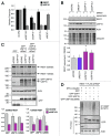
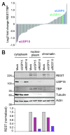
Figure 1. A screen for DUBs that modulate the amount of REST in lung cancer cells identifies USP15. (A) Identification of USP15 from an unbiased DUB siRNA screen. A549 cells were transfected with REST siRNA or a library of pooled siRNA oligos _targeting 85 human DUBs. REST levels in the nucleoplasmic fraction were determined 72 h later by immunoblotting and normalized to TATA binding protein (TBP). The candidates for stabilization of REST were USP49, USP47, USP15 and USP52, in contrast to the USP15 paralogs USP4 and USP11. (B). USP15 depletion reduces the amount of REST without influencing its subcellular localization. Following transfection with siRNA _targeting REST (siREST-5), pooled siRNAs _targeting USP15 or no oligonucleotide (mock), A549 cells were fractionated into cytoplasm, nucleoplasm and a pellet containing chromatin. Extracts were immunoblotted and probed for REST and USP15 (monoclonal); tubulin and TBP were used as cytoplasmic and nucleoplasmic markers, respectively. Quantification of REST normalized to actin, which was detected in each fraction, is shown below.
None
Figure 2. REST abundance is dependent on the amount of cellular USP15. (A) Multiple siRNAs for USP15 but not USP4 or USP11 reduce the amount of REST. siRNA-mediated knockdown was repeated with five individual USP15 oligos and compared with pooled siRNAs _targeting USP4 and USP11. After 72 h, proteins were prepared from subcellular fractions and immunoblotted. Representative blots for REST in the nucleoplasmic fraction are shown with mean quantification normalized to actin and the appropriate siRNA control below (n = 3 individual experiments, error bars show standard deviation). (B) Confirmation of specificity for siRNAs _targeting the USP15 paralogs. Cytoplasmic protein extracts from the experiments shown in (A) were immunoblotted for USP15 (polyclonal, *indicates cross-reactivity with USP11), USP4 and USP11 to evaluate knockdown efficiency. (C) The amount of REST correlates with USP15 levels. A scatter plot representing the mean quantification of REST (x-axis) plotted against that of USP15 (y-axis) for each of the controls or samples treated with siRNA _targeting USP15, USP4 or USP11 in these experiments; linear regression R2 = 0.69.
None
Figure 3. USP15 can exert a post-translational effect on REST. (A) The effect of USP15 depletion on REST protein abundance is not predicated on reduction of REST mRNA. A549 cells were transfected with siRNAs as indicated and RNA was prepared 72 h later. REST or USP15 transcripts were quantified relative to β-actin (ACTB) by qRT-PCR; the mean of four independent experiments is shown (bars show standard error). (B) The loss of REST in USP15-depleted cells can be rescued by proteasome inhibition. A549 cells were transfected with siRNA as indicated for 72 h and treated with 50 nM epoxomicin for the final 6 h prior to whole-cell lysis and immunoblotting. Representative blots are shown with the mean fold induction of REST following proteasome inhibition plotted below [siUSP15-1, n = 5, *p = 0.045 (epoxomicin/DMSO); siUSP15-2, n = 3, p = 0.098; bars show standard error]. (C) Catalytically active USP15 can partially rescue levels of nascent REST. HEK-293T cells were transfected with the indicated plasmids 24 h after treatment with USP15 siRNA or control reagents. Whole-cell protein extracts were prepared 48 h later for immunoblotting. The amount of unglycosylated 120 kDa REST or O-glycosylated 220 kDa REST were determined from three independent experiments and normalized to actin; all values are expressed relative to cells transfected with control siRNA and a plasmid expressing GFP alone (lane 1) (n = 3, error bars show standard deviation). (D) Catalytic activity is required for USP15 to reverse ubiquitylation of REST. HEK-293T cells were transfected with the indicated constructs for 48 h and treated with epoxomicin for the last 6 h, before immunoprecipitation with an anti-REST antibody and immunoblotting for myc-tagged ubiquitin. A representative blot from two independent experiments is shown.
None
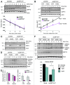
Figure 4. USP15 primarily stabilizes newly synthesized REST. (A) USP15 depletion does not accelerate REST turnover in the absence of protein synthesis. A549 cells were transfected with siRNA as indicated and 68 h later cycloheximide (CHX) was added. Cells were sampled over 4 h, and whole-cell protein extracts were analyzed by immunoblotting. Quantification of REST normalized to actin is shown below for three independent experiments; the half-life under control conditions is indicated by the dotted line. Mean values for REST are normalized to the level before CHX treatment; error bars show standard deviation (siCON2: R2 = 0.926, gradient -0.208; siUSP15-P: R2 = 0.934, gradient -0.201). (B) USP15 depletion impairs accumulation of newly translated REST. A549 cells were transfected with siRNA, and after 68 h (−4 h) treated with cycloheximide for 4 h, before washing the cells and releasing into fresh medium (0 h). Whole-cell protein extracts were sampled and analyzed by immunoblotting. Quantification of 220 kDa REST normalized to actin is shown below for three independent experiments. Mean values for REST are normalized to the maximum recovery (3 h with siCON2); error bars show standard deviation (siCON2: R2 = 0.996, gradient 0.266; siUSP15-P: R2 = 0.990, gradient 0.159). (C) USP15 depletion does not generically block translational recovery. A549 cells were transfected and treated as in (B); whole-cell extracts were probed for NRF2. (D) USP15 depletion impairs accumulation of both nascent (120 kDa) and glycosylated (220 kDa) REST on cycloheximide washout. A549 cells were transfected with siRNA and treated with cycloheximide and analyzed as described in (B). (E) The effect of USP15 on accumulation of nascent REST is not due to altered REST mRNA. A549 cells were transfected and treated as in (B); RNA was prepared and transcript levels were determined relative to ACTB by QPCR. Data are shown normalized to mock-transfected cells prior to cycloheximide treatment (n = 3 independent experiments, error bars show standard deviation). (F) USP15 exerts a post-translational effect on newly synthesized REST. A549 cells were transfected with siRNA and after 68 h (−4 h) were treated with cycloheximide for 4 h before washing the cells and releasing into fresh medium containing 50 nM epoxomicin. Whole-cell protein extracts were sampled and analyzed by immunoblotting. A non-specific band is indicated by an asterisk (*ns). Quantification of 120 kDa REST normalized to actin is shown below, 3 h into recovery from the translational block in the absence or presence of epoxomicin (n = 3 independent experiments, error bars show standard deviation, *p < 0.05).
None
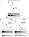
Figure 5. A small fraction of endogenous USP15 associates with polysomes. (A) USP15 co-fractionates with polysomes. HEK-293T cells were subject to gradient density centrifugation and fractions immunoblotted for the small ribosomal subunit protein RPS6, the soluble protein GAPDH and USP15. The A254nm profile was used to determine the fractions containing polysomes, free ribosomes and ribosomal subunits. (B) Dissociation of polysomes to 80S ribosomes shifts USP15 but not GAPDH within the density gradient. The experiment in (A) was repeated including a replicate sample that was treated with RNase for 1 min prior to gradient density centrifugation.
None
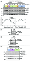
Figure 6. REST is degraded at mitosis and resynthesised as cells enter G1. (A) REST accumulates on release from G1/S arrest and degrades in cells arrested at G2/M. A549 cells were synchronized with thymidine and released from G1/S into media (left) or media containing nocodazole (right). Protein expression was monitored by immunoblotting over 14 h. Total REST and USP15 were quantified from duplicate gels, normalized to actin and plotted relative to the level before release from the thymidine block. (B and C) Phosphorylated forms of both REST (B) and USP15 (C) accumulate during prometaphase arrest. Thymidine synchronized A549 cells were released into nocodazole for 12 h and lysed with E1A buffer. Protein extract was incubated in buffer alone (−) or with lambda protein phosphatase (LPP, +) prior to immunoblotting for REST or USP15; untreated extract (input) is shown for comparison. P-USP15 is indicated with an arrow, above the USP15 doublet seen in asynchronous cells. (D) Phosphorylated REST degrades at mitosis and unphosphorylated REST accumulates as cells enter G1. A549 cells were synchronized with thymidine and then arrested in nocodazole for 14 h before shaking off the mitotic cells and releasing them from G2/M arrest (0 h) into fresh medium. Protein expression was monitored by immunoblotting.
None
Figure 7. USP15 is required for the recovery of REST levels on mitotic exit. (A) USP15 depletion does not prevent degradation of phosphorylated REST at mitosis but limits its recovery. A549 were transfected with siRNA prior to thymidine synchronization and then arrested in nocodazole for 14 h before shaking off the mitotic cells and releasing from G2/M arrest (0 h). Protein expression was monitored for 4 h, and a representative immunoblot is shown. (B) USP15 depletion impairs recovery of REST at mitotic exit. Cells were treated as described in (A) and extracts collected for immunoblotting before release from G2/M (0 h) or in early G1 (4 h). Mean quantification of REST normalized to actin is plotted, relative to that in asynchronous cells, for four independent experiments (bars show standard error, **p = 0.0005). (C) Individual USP15 siRNAs recapitulate the block on REST accumulation in early G1. Cells were transfected with siRNAs as indicated, then treated and extracts collected as in (B). A representative immunoblot from two independent experiments is shown at 4 h post-release from nocodazole. (D) A model for REST regulation by USP15. USP15 does not oppose the phosphorylation-dependent eradication of REST at mitosis (M). Instead, during G1 and G2, USP15 counteracts ubiquitylation of newly synthesized REST, allowing its levels to accumulate.

Comment in

Similar articles

Cited by

References

    1. Hammond-Martel I, Yu H, Affar B. Roles of ubiquitin signaling in transcription regulation. Cell Signal. 2012;24:410–21. doi: 10.1016/j.cellsig.2011.10.009. - DOI - PubMed
    1. Clague MJ, Coulson JM, Urbé S. Cellular functions of the DUBs. J Cell Sci. 2012;125:277–86. doi: 10.1242/jcs.090985. - DOI - PubMed
    1. Grabbe C, Husnjak K, Dikic I. The spatial and temporal organization of ubiquitin networks. Nat Rev Mol Cell Biol. 2011;12:295–307. doi: 10.1038/nrm3099. - DOI - PMC - PubMed
    1. Komander D, Clague MJ, Urbé S. Breaking the chains: structure and function of the deubiquitinases. Nat Rev Mol Cell Biol. 2009;10:550–63. doi: 10.1038/nrm2731. - DOI - PubMed
    1. Sacco JJ, Coulson JM, Clague MJ, Urbé S. Emerging roles of deubiquitinases in cancer-associated pathways. IUBMB Life. 2010;62:140–57. - PMC - PubMed

Publication types

  NODES
twitter 2