. 2013 May 29;5(187):187ra69.
doi: 10.1126/scitranslmed.3005066.

Coronary microvascular pericytes are the cellular _target of sunitinib malate-induced cardiotoxicity

Affiliations

Coronary microvascular pericytes are the cellular _target of sunitinib malate-induced cardiotoxicity

Vishnu Chintalgattu et al. Sci Transl Med. .

Abstract

Sunitinib malate is a multi_targeted receptor tyrosine kinase inhibitor used in the treatment of human malignancies. A substantial number of sunitinib-treated patients develop cardiac dysfunction, but the mechanism of sunitinib-induced cardiotoxicity is poorly understood. We show that mice treated with sunitinib develop cardiac and coronary microvascular dysfunction and exhibit an impaired cardiac response to stress. The physiological changes caused by treatment with sunitinib are accompanied by a substantial depletion of coronary microvascular pericytes. Pericytes are a cell type that is dependent on intact platelet-derived growth factor receptor (PDGFR) signaling but whose role in the heart is poorly defined. Sunitinib-induced pericyte depletion and coronary microvascular dysfunction are recapitulated by CP-673451, a structurally distinct PDGFR inhibitor, confirming the role of PDGFR in pericyte survival. Thalidomide, an anticancer agent that is known to exert beneficial effects on pericyte survival and function, prevents sunitinib-induced pericyte cell death in vitro and prevents sunitinib-induced cardiotoxicity in vivo in a mouse model. Our findings suggest that pericytes are the primary cellular _target of sunitinib-induced cardiotoxicity and reveal the pericyte as a cell type of concern in the regulation of coronary microvascular function. Furthermore, our data provide preliminary evidence that thalidomide may prevent cardiotoxicity in sunitinib-treated cancer patients.


Figures

Fig. 1
Fig. 1. Sunitinib treatment induces coronary microvascular dysfunction
(A) Representative cardiac MRI images from mice treated with sunitinib or vehicle for 14 days. (B) Left ventricular ejection fraction of treated mice at various time points (n= 7–10 mice per group). (C) Heart weight/body weight and lung weight/body weight ratios of mice treated for 14 days (n=7–10 mice per group). (D) Mean arterial pressure (MAP) in sunitinib or vehicle-exposed mice measured by tail cuff (n=5–6 mice per group). (E) Representative ultrasound tracings of dilated (induced with 2.5% isoflurane) and basal (with 1% isoflurane) coronary flow after 14 days of treatment. (F) Quantification of coronary flow reserve (dilated/basal flow) in sunitinib-treated and vehicle-treated mice (n=6–10 mice per group). (G) Total coronary flow in response to increasing concentrations of adenosine measured in ex-vivo heart preparations from sunitinib or vehicle-treated mice. (H) Quantitative RT-PCR results probing for PHD3 (normalized to GAPDH) using total RNA extracted from cardiac tissue of mice treated for the indicated number of days. * p <0.05
Fig. 2
Fig. 2. Sunitinib augments pressure overload-induced cardiac dysfunction
(A) Cardiac contractile reserve (maximal developed pressure over time, dP/dtmax) in response to increasing concentrations of dobutamine in sunitinib or vehicle-treated mice (n=7–8). (B) Heart weight/body weight and lung weight/body weight ratios of sunitinib or vehicle-treated mice (n=8–9) after 14 days of TAC. (C) Cardiac ejection fraction in sunitinib or vehicle-treated mice as measured by cardiac MRI after 0, 7 and 14 days of TAC (n=9–10). (D) Coronary flow reserve in sunitinib or vehicle-treated mice after 0, 7, and 14 days of TAC (n=7–10). (E) Quantification of vessel densities (CD31+ vessels/cardiomyocyte) in cardiac sections from sunitinib or vehicle-treated mice after 14 days of TAC or 14 days of treatment without TAC stress. (F) Immunohistochemical staining for CD31 in cardiac sections from sunitinib or vehicle-treated mice after 14 days of TAC. Scale bar = 50 µm. (G) Representative photomicrographs of Masson’s trichrome stained cardiac sections from sunitinib or vehicle-treated mice after 14 days of TAC. Scale bar = 50 µm. (H) Phosphorylated and total PDGFR β protein in pre-TAC and 14-day TAC mice treated with sunitinib or vehicle. All results presented in this figure are representative of 2–3 independent experiments. * p < 0.05
Fig. 3
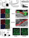
Fig. 3. Coronary microvascular dysfunction in sunitinib-treated mice is associated with loss of pericytes
(A) Coronary flow reserve in response to acetylcholine in mice treated for 14 days. (B) Total coronary flow in response to increasing concentrations of sodium nitroprusside measured ex vivo using hearts from sunitinib-treated or control mice after 14 days (n=7–8 hearts per group). (C) NG2 protein expression in cardiac lysates from mice treated for 14 days. (D) Densitometric quantification (from Western blotting) of NG2 protein normalized to GAPDH at 3, 7, 14, or 21 days in hearts from sunitinib-treated mice relative to controls (n=4–5 mice per time point); p-value calculated by ANOVA. (E) Confocal micrographs from representative cardiac sections showing staining for the endothelial cell marker CD31 (red), the pericyte marker NG2 (green), nuclear staining with DAPI (blue) and the merged image (bottom right) from sunitinib (bottom panels) or vehicle-treated mice (top panels) after 14 days. Scale bar = 20 µm. (F) Quantification of NG2+ pericyte coverage of CD31+ microvessels in sunitinib or vehicle-treated mice after 14 or 21 days of treatment (n=4 animals per group). (G) Representative scanning electron micrographs of the cardiac microvasculature from control or sunitinib-treated mice after 14 days of treatment. Pseudocoloring is used (green for pericytes, red for microvessels) to aid in distinguishing pericytes from underlying microvessels; gray-scale electron micrographs are shown in Fig. S2E. Scale bar =5 µm. (H) Representative images of the coronary microvasculature derived from computer-based reconstruction of dextran-perfused hearts of sunitinib-treated mice (bottom panel) compared with vehicle-treated mice (top panel). * p < 0.05.
Fig. 4
Fig. 4. Pericyte abnormalities are not observed in skeletal muscle or after doxorubicin treatment
Percent contraction or relaxation in the presence of (A) phenylephrine (PE) or (B) acetylcholine (Ach), respectively, in thoracic aortas isolated from mice treated with either sunitinib or vehicle (n=4–5). (C) Confocal micrographs from representative skeletal muscle (gastrocnemius) sections showing staining for the endothelial cell marker CD31 (red), the pericyte marker NG2 (green), nuclear staining with DAPI (blue) and the merged image (bottom right) from sunitinib (right panels) or vehicle-treated mice (left panels) after 14 days. Scale bar = 20 µm. (D) Quantification of coverage of CD31+ microvessels by NG2+ pericytes in heart and skeletal muscle isolated from treated mice (n=4 mice per group). (E) Cardiac ejection fraction in doxorubicin or vehicle-treated mice, as measured by cardiac MRI (n=4–5 mice per group). (F) Coronary flow reserve in doxorubicin or vehicle-treated mice (n=4–5 mice per group). (G) NG2 protein expression (by Western blot) in cardiac lysates from doxorubicin or vehicle-treated mice. (H) Confocal micrographs from representative cardiac sections showing staining for the endothelial cell marker CD31 (red), pericyte marker NG2 (green), nuclear stain for DAPI (blue) and the merged image (bottom right) from mice treated with vehicle control (left panels), 5 mg/kg/wk doxorubicin (center panels) or 10 mg/kg/wk doxorubicin (right panels). Scale bar = 20 µm. All results presented in this figure are representative of 2–3 independent experiments, * p < 0.05.
Fig. 5
Fig. 5. Sunitinib-induced coronary dysfunction and pericyte loss is recapitulated by a PDGFR inhibitor
(A) Chemical structures of sunitinib and CP-673451. (B) Heart weight or lung weight to body weight ratios in CP-673451 or vehicle-treated mice (n=5 mice per group). (C) Left ventricular ejection fractions assessed at seven or 14 days after initiation of treatment with either CP-673451 or vehicle control as measured by cardiac MRI (n=5 mice per group). (D) Coronary flow reserve assessed at seven or 14 days after initiation of treatment with CP-673451 or vehicle (n=5 mice per group). (E) Confocal micrographs from representative cardiac sections showing staining for CD31 (red), NG2 (green), and the merged image (bottom left) from mice treated with vehicle control (top panels) or CP-673451 for 14 days. Scale bar = 20 µm. (F) Western blot probed for NG2 and phospho-/total levels of PDGFR-β from hearts of mice treated with vehicle for seven days or with CP-673451 for three or seven days. Concentrations of PDGFR-β (G) or NG2 (H) in cardiac lysates from mice treated with CP-673451 for three or seven days relative to levels in control-treated mice, as measured by quantitative densitometry. Values are normalized to levels of GAPDH (n=4 independent samples per group). * p < 0.05
Fig. 6
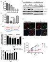
Fig. 6. Sunitinib-induced pericyte cytotoxicity is ameliorated by co-treatment with thalidomide
(B) Pericyte viability measured by MTT assay after incubation for 24 hours with sunitinib at the indicated dosages. 0 nM sunitinib cells were treated with DMSO only, and data are presented as MTT values relative to 0 nM sunitinib cell values. (B) Pericyte viability measured by MTT assay after 24 hours of treatment with DMSO (control), sunitinib malate (SM, 1 µM), thalidomide (Thal, 1 µM) plus sunitinib (SM + Thal) or thalidomide alone (Thal). Data presented as MTT values relative to control. (C) Overview of the protocol for in vivo thalidomide rescue experiments. (D) Coronary flow reserve in sunitinib-treated mice co-treated with either vehicle or thalidomide for 14 days followed by a 14-day recovery period (n=5–8 mice per group per time point). Data are shown as coronary flow reserve index, the ratio of coronary flow reserve in mice treated with sunitinib plus vehicle to coronary flow reserve in mice treated with sunitinib plus thalidomide (denoted “vehicle/thalidomide”). (E) Left ventricular ejection fraction in sunitinib-treated mice co-treated with either vehicle or thalidomide for 14 days followed by a 14-day recovery period. (F) Western blot of cardiac lysates from mice co-treated with sunitinib plus vehicle or thalidomide after 14 days of treatment (left) or after recovery for 14 days, probed for NG2 and phospho-/total PDGFR-β. (G) Confocal micrographs from representative cardiac sections showing staining for CD31 (red), NG2 (green), DAPI (bottom left) and the merged image (bottom right) from mice treated with sunitinib + vehicle (left panels) or sunitinib + thalidomide (right panels) for 14 days. Scale bar = 20 µm. (H) Average tumor volume over time in vehicle, thalidomide, sunitinib or sunitinib + thalidomide treated mice (n=6–10 mice per group). All results presented in this figure are representative of 2–4 independent experiments, * p < 0.05.

Comment in

Similar articles

Cited by

References

    1. Di Lorenzo G, Autorino R, Bruni G, Carteni G, Ricevuto E, Tudini M, Ficorella C, Romano C, Aieta M, Giordano A, Giuliano M, Gonnella A, De Nunzio C, Rizzo M, Montesarchio V, Ewer M, De Placido S. Cardiovascular toxicity following sunitinib therapy in metastatic renal cell carcinoma: a multicenter analysis. Annals of Oncology. 2009 Sep;20:1535. - PubMed
    1. Schmidinger M, Zielinski CC, Vogl UM, Bojic A, Bojic M, Schukro C, Ruhsam M, Hejna M, Schmidinger H. Cardiac toxicity of sunitinib and sorafenib in patients with metastatic renal cell carcinoma. Journal of clinical oncology : official journal of the American Society of Clinical Oncology. 2008 Nov 10;26:5204. - PubMed
    1. Khakoo AY, Kassiotis CM, Tannir N, Plana JC, Halushka M, Bickford C, 2nd, Trent J, Champion JC, Durand JB, Lenihan DJ. Heart failure associated with sunitinib malate: a multi_targeted receptor tyrosine kinase inhibitor. Cancer. 2008 Jun;112:2500. - PubMed
    1. Chu TF, Rupnick MA, Kerkela R, Dallabrida SM, Zurakowski D, Nguyen L, Woulfe K, Pravda E, Cassiola F, Desai J, George S, Morgan JA, Harris DM, Ismail NS, Chen JH, Schoen FJ, Van den Abbeele AD, Demetri GD, Force T, Chen MH. Cardiotoxicity associated with tyrosine kinase inhibitor sunitinib. Lancet. 2007 Dec 15;370:2011. - PMC - PubMed
    1. Richards CJ, Je Y, Schutz FA, Heng DY, Dallabrida SM, Moslehi JJ, Choueiri TK. Incidence and risk of congestive heart failure in patients with renal and nonrenal cell carcinoma treated with sunitinib. Journal of clinical oncology : official journal of the American Society of Clinical Oncology. 2011 Sep 1;29:3450. - PubMed

Publication types

  NODES
Note 1
twitter 2