Review
. 2013 Oct;11(10):1141-58.
doi: 10.1158/1541-7786.MCR-13-0244. Epub 2013 Aug 8.

Cancer gene discovery: exploiting insertional mutagenesis

Affiliations
Review

Cancer gene discovery: exploiting insertional mutagenesis

Marco Ranzani et al. Mol Cancer Res. 2013 Oct.

Abstract

Insertional mutagenesis has been used as a functional forward genetics screen for the identification of novel genes involved in the pathogenesis of human cancers. Different insertional mutagens have been successfully used to reveal new cancer genes. For example, retroviruses are integrating viruses with the capacity to induce the deregulation of genes in the neighborhood of the insertion site. Retroviruses have been used for more than 30 years to identify cancer genes in the hematopoietic system and mammary gland. Similarly, another tool that has revolutionized cancer gene discovery is the cut-and-paste transposons. These DNA elements have been engineered to contain strong promoters and stop cassettes that may function to perturb gene expression upon integration proximal to genes. In addition, complex mouse models characterized by tissue-restricted activity of transposons have been developed to identify oncogenes and tumor suppressor genes that control the development of a wide range of solid tumor types, extending beyond those tissues accessible using retrovirus-based approaches. Most recently, lentiviral vectors have appeared on the scene for use in cancer gene screens. Lentiviral vectors are replication-defective integrating vectors that have the advantage of being able to infect nondividing cells, in a wide range of cell types and tissues. In this review, we describe the various insertional mutagens focusing on their advantages/limitations, and we discuss the new and promising tools that will improve the insertional mutagenesis screens of the future.


Figures

Figure 1
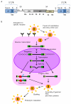
Figure 1. General outline of the work-flow for cancer gene discovery using insertional mutagens
From top-left, clockwise: the insertional mutagens are administered or activated in the proper mouse model to induce tumorigenesis. Integrations are retrieved from tumor DNA by a PCR-based technique and sequenced. Bioinformatic and statistical analysis leads to map vector integrations events. Clusters of these events are called common insertion sites (CIS). The candidate cancer genes hosted within CIS are then validated by in vitro and/or in vivo experiments for their transformation potential. The orthologs are then interrogated for their expression level and mutation status in human tumors, allowing the identification of candidate therapeutic _targets for the human disease. RE: restriction enzyme.
Figure 2
Figure 2. Different mechanisms of gene deregulation by insertional mutagenesis
Vector integration can induce insertional mutagenesis by different mechanisms: A) enhancer insertions occur when the enhancer elements of the integrating vector enhance the transcription of a cellular gene under its own promoter. The integrated vector might be some distance from the cellular gene, and usually is found upstream of the gene in antisense orientation. B) Promoter insertions occur when the mutagen integrates in the sense orientation of a gene, and uncouples the gene transcription from its own promoter by placing it under the control of the vector one. A chimeric transcript encoding the full-length protein can be thus generated. C) Landing of a vector within the transcriptional unit of a gene (intragenic insertions) can induce the generation of a truncated transcript via the internal polyA signal (upper panel) or disrupt an exon (lower panel). If a chimeric transcript encoding a truncated version of the cellular protein is generated, the new protein might have an abnormal biological activity (e.g. gain-of-function of a proto-oncogene). Alternatively, the intragenic vector might disrupt the normal transcription and impede the correct translation of the cellular protein (e.g. loss-of-function of a TSG). In the three boxes, the vector is depicted in purple, with enhancer/promoter (E/P) elements in green. Splice donors (SD), splice acceptors (SA) and polyadenylation sites (pA) are indicated. A prototypical cellular gene is shown as a sequence of three boxes corresponding to three exons, with orange boxes indicating protein-coding exons and green segments indicating untranslated regions.
Figure 3
Figure 3. Structure of the genome and replication life cycle of retroviruses
A) Schematic representation of the proviral form of a retrovirus with its simple genomic architecture. The coding sequences of the three essential retroviral genes, gag, pol, and env, and their relative protein subunits are indicated below. EP: enhancer-promoter; att: attachment site; TSS: transcription start site; pA: polyadenylation signal; PBS: primer binding site, necessary for the binding of the primer from which the retrotranscription is triggered; SD: splice donor; ψ: viral packaging signal; SA: splice acceptor; PPT: polypurine tract; SU: surface; TM: transmembrane; RT: retrotranscriptase; IN: integrase; NC: nucleocapsid; CA: capsid; PR: protease; MA: matrix. Modified from (113). B) Retroviral replication cycle. The parental virus attaches to a specific receptor on the surface of a susceptible cell with the SU portion of the viral Env protein leading to fusion and entry of the core. Reverse transcription then generates a double-stranded DNA copy of the RNA genome. The provirus is transported into the nucleus and integrated into chromosomal DNA. It is then transcribed by cellular RNA polymerase II. Transcription generates RNA copies with the terminal structures organized as in the parental genome. These copies become full-length and spliced messenger RNAs as well as full-length progeny virion RNAs. Viral messages are translated in the cytoplasm. Virion proteins and progeny RNAs assemble at the cell periphery and the plasma membrane, and progeny viruses are released by a process of budding and subsequent maturation into infectious viruses. Modified from (9).
Figure 4
Figure 4. Sleeping Beauty structure and mechanism of transposition
(A) The Sleeping Beauty (SB) inverted terminal repeats (light blue arrows) each contain two direct repeats (DRs) (violet and dark blue triangles) that are the binding sites for the transposase. The genomic TA dinucleotide that is duplicated during integration and flanks the transposon is shown in red. (B) Cut and paste mechanism of SB transposition. The transposase (green triangles) initiates transposition by cleaving both ends of the transposon to generate 3 base pair (bp) 5′ overhangs (violet) and also cleaves a genomic TA dinucleotide at the integration site (pink) to create a gap with a 3′ TA overhang at both ends. The host non-homologous end joining (NHEJ) DNA repair machinery then repairs the single-stranded gaps at the integration site and the double-strand breaks in the donor DNA. A small 5 bp footprint (one TA and 3 bps from the end of the transposon) remains at the excision site.
Figure 5
Figure 5. Transposons for insertional mutagenesis
A) Sleeping Beauty T2/Onc2 can deregulate the expression of oncogenes or inactivate the expression of tumor suppressor genes. T2/Onc2 contains a murine stem cell virus (MSCV) 5′ long terminal repeat (LTR) and a splice donor (SD) site derived from exon 1 of the mouse Foxf2 gene. T2/Onc2 can thus promote the expression of an oncogene when integrated upstream of or within the gene in the same transcriptional orientation. T2/Onc2 is flanked by optimized SB transposase binding sites (violet and dark blue triangles) that are located within the transposon inverted terminal repeats (light blue arrows), which increase the frequency of SB transposition (upper panel). T2/Onc2 also contains two splice acceptors and a bi-directional polyA (pA) and can thus prematurely terminate transcription of a tumor suppressor gene when integrated within the tumor suppressor gene in either orientation (pale blue). One splice acceptor is derived from exon 2 of mouse engrailed 2 (En2-SA) and the other from the Carp b-actin gene (SA) (lower panel). B) The PB-based insertional mutagenesis system described in (63). Mouse lines carrying the genetic components of the transposon systems. Upper Panel: RosaPB and RosaSB knock-in mice express PiggyBac or Sleeping Beauty transposase under control of the constitutively active Rosa26 promoter. Lower panel: Transposon design and transposon mouse lines. Three transposon constructs were designed, which differ in their promoter and enhancer. All three transposons have PB as well as SB IR/DRs and can therefore be mobilized with both transposases. CβASA, Carp β-actin splice acceptor; En2SA, Engrailed-2 exon-2 splice acceptor; SD, Foxf2 exon-1 splice donor; pA, bidirectional SV40 polyadenylation signal; CAG, cytomegalovirus enhancer and chicken beta-actin promoter; MSCV, murine stem cell virus long terminal repeat; PGK, phosphoglycerate kinase promoter. Modified from (8)
Figure 6
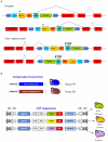
Figure 6. Four plasmids system for the production of late generation LVs
Top panels: LV packaging constructs encoding HIV structural proteins (gag/pro/pol), Envelope construct (Env) encoding for the envelope glycoprotein and Rev. Lower panel: LV transfer vector; the HIV 5′LTR U3 has been substituted with Cytomegalovirus promoter-enhancer sequence; the 3′LTR U3 sequence containing the HIV promoter-enhancer region has been deleted. Upon retrotranscription the 3′LTR is copied at the 5′ obtaining a Self-Inactivating (SIN) vector. Wpre: Woodchuck post-transcriptional regulatory element; CMV: cytomegalovirus enhancer/promoter sequence; SD: splice donor; SA: splice acceptor; ψ: packaging signal.

Similar articles

Cited by

References

    1. Selten G, Cuypers HT, Zijlstra M, Melief C, Berns A. Involvement of c-myc in MuLV-induced T cell lymphomas in mice: frequency and mechanisms of activation. The EMBO journal. 1984;3:3215–22. - PMC - PubMed
    1. Uren AG, Kool J, Berns A, van Lohuizen M. Retroviral insertional mutagenesis: past, present and future. Oncogene. 2005;24:7656–72. - PubMed
    1. Morishita K, Parker DS, Mucenski ML, Jenkins NA, Copeland NG, Ihle JN. Retroviral activation of a novel gene encoding a zinc finger protein in IL-3-dependent myeloid leukemia cell lines. Cell. 1988;54:831–40. - PubMed
    1. van Lohuizen M, Breuer M, Berns A. N-myc is frequently activated by proviral insertion in MuLV-induced T cell lymphomas. The EMBO journal. 1989;8:133–6. - PMC - PubMed
    1. Kool J, Berns A. High-throughput insertional mutagenesis screens in mice to identify oncogenic networks. Nature reviews Cancer. 2009;9:389–99. - PubMed

Publication types

Substances

  NODES
admin 1
INTERN 1
twitter 2