. 2013 Sep 1;73(17):5556-68.
doi: 10.1158/0008-5472.CAN-13-0013. Epub 2013 Aug 16.

Hbo1 is a cyclin E/CDK2 substrate that enriches breast cancer stem-like cells

Affiliations

Hbo1 is a cyclin E/CDK2 substrate that enriches breast cancer stem-like cells

Mylinh T Duong et al. Cancer Res. .

Abstract

Expression of cyclin E proteolytic cleavage products, low-molecular weight cyclin E (LMW-E), is associated with poor clinical outcome in patients with breast cancer and it enhances tumorigenecity in mouse models. Here we report that LMW-E expression in human mammary epithelial cells induces an epithelial-to-mesenchymal transition phenotype, increases the CD44(hi)/CD24(lo) population, enhances mammosphere formation, and upregulates aldehyde dehydrogenase expression and activity. We also report that breast tumors expressing LMW-E have a higher proportion of CD44(hi)/CD24(lo) tumor cells as compared with tumors expressing only full-length cyclin E. In order to explore how LMW-E enriches cancer stem cells in breast tumors, we conducted a protein microarray analysis that identified the histone acetyltransferase (HAT) Hbo1 as a novel cyclin E/CDK2 substrate. The LMW-E/CDK2 complex phosphorylated Hbo1 at T88 without affecting its HAT activity. When coexpressed with LMW-E/CDK2, wild-type Hbo1 promoted enrichment of cancer stem-like cells (CSC), whereas the T88 Hbo1 mutant reversed the CSC phenotype. Finally, doxorubicin and salinomycin (a CSC-selective cytotoxic agent) synergized to kill cells expressing LMW-E, but not full-length cyclin E. Collectively, our results suggest that the heightened oncogenecity of LMW-E relates to its ability to promote CSC properties, supporting the design of therapeutic strategies to _target this unique function.


Conflict of interest statement

The authors disclose no potential conflicts of interest.

Figures

Figure 1
Figure 1. LMW-E activates gene expression associated with the EMT
76NE6 cells with stable expression for cyclin E (V, EL, LMW-E) and 3 of the in vivo passaged lines (T1G2.2, T1G3.1, T1G4.2) were (A) fixed and immunostained with E-cadherin antibody and nuclei were counterstained with DAPI (scale bar = 100 μm), and (B) subjected to western blot analysis with cyclin E and E-cadherin. (C – H) Cells were cultured on monolayer culture to 70% confluency and RNA were extracted and subjected to qRT-PCR analysis for the mRNA levels of cyclin E (C), E-cadherin (D), twist (E), vimentin (F), N-cadherin (G), and slug (H). These values were normalized against GAPDH mRNA levels and statistical analysis used was unpaired student's t-test (error bars = +/- SEM).
Figure 2
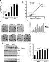
Figure 2. LMW-E expression enriches for the mammary CSC population
(A) 76NE6 cells with stable expression for cyclin E (V, EL, LMW-E) and 3 of the in vivo passaged lines (T1G2.2, T1G3.1, T1G4.2) were incubated with PE-CD24 and APC-CD44 antibodies and analyzed with a FACS Calibur. Statistical analysis used was unpaired student's t-test (error bars = +/- SEM). (B) The percentage of CD44hi/CD24lo population of these cells were subjected to linear correlation analysis versus cyclin E mRNA levels (error bars = +/- SEM). (C) Representative bright field images of cells cultured on ultra-low attachment plates for 7 days (scale bar =100 μm). The vector control cells undergo apoptosis as apparent by extensive membrane blebbing (white arrows). (D) MTT solution was added to the mammosphere cultures at 500 μg/ml concentration and incubated at 37°C for 1 hour. Number of mammospheres were quantified using the automated GelCount program and statistical analysis used was unpaired student's t-test (error bars = +/- SEM). For secondary mammosphere assay, cells from primary mammospheres were dispersed with 0.05% trypsin, seeded in 6-well ultra-low attachment plates in mammosphere media and incubated for 5 days and counted as described above. (E) Lysates from cell cultured on regular tissue culture treated plates were extracted and subjected to Western blot analysis with antibodies against cyclin E, CD24, CD44 and β-actin. Additionally, lysates from mammosphere cultures were extracted and subjected to Western blot analysis with antibodies against ALDH and β-actin. (F) ALDELUOR assay was performed on cells cultured on monolayer. For each sample, 500,000 cells were incubated with the ALDH substrate alone or with the ALDH inhibitor (DEAB) and subjected to FACS analysis. The ALDH activity is obtained by subtracting the DEAB inhibitor value and the statistical analysis used was unpaired student's t-test (error bars = +/- SEM).
Figure 3
Figure 3. Hbo1 is a novel substrate of the cyclin E/CDK2 complex
(A) The ProtoArray microarray experiment was performed in two independent experiments. The phosphorylation signals indicate relative radioactive signal detected in the microarray spots. (B) FLAG-EL or LMW-E, HA-CDK2, and Myc-Hbo1 constructs were transfected into HEK293T cells and IP with either Myc-tagged or FLAG-tagged antibodies and probed with the indicated antibodies. (C) SFB-EL or SFB-LMW-E was co-transfected with SFB-CDK2 into HEK293T cells, purified using FLAG-tagged antibody, eluted with 3X FLAG peptide and visualized by Western blot analysis. Myc-Hbo1 was transfected into HEK293T cells and purified using Myc-tagged antibody (D) The EL/CDK2 or LMW-E/CDK2 kinase complex was incubated with purified Hbo1 in the presence of 32P-γ-ATP, with or without roscovitine. The samples were separated by SDS-PAGE and exposed to x-ray films. (E) Schematic of the Hbo1 gene construct with the potential phosphorylation sites predicted based on the CDK2 consensus phosphorylation motif (S/T-P-X-R/K/H). (F & G) The six potential phosphorylation sites were mutated to alanine, expressed, purified and subjected to similar kinase assay as in (D).
Figure 4
Figure 4. Hbo1 is overexpressed in breast cancer cell lines and co-expression with LMW-E/CDK2 enhances self-renewal capability of hMECs
(A) Cell lysates from 3 hMECs lines and 18 breast cancer cell lines were subjected to Western blot analysis with antibodies to Hbo1 and β-actin. (B) The 76NE6 stable cell panel and the TDCs were subjected to similar analysis as in (A). (C) Lentivirus generated in HEK293T cells and carrying the EL, LMW-E, CDK2, or Hbo1 (wt, T88A, or T88D) constructs were used to infect the 76NE6 cells and then subjected to mammosphere culture. The results were averaged from at least 2 independent experiments and the statistical analysis used was unpaired student's t-test (error bars = +/- SEM). (D-G) shControl, shHbo1-4, and shHbo1-5 lentivirus were produced in HEK293T cells and used to infect the 76NE6 stable cell panel, two of the TDCs and Sum159 cells and subjected to (D) GFP FACS analysis, (E) Western blot analysis, (F) CD44/CD24 FACS analysis, and (G) mammosphere formation assay. These cells were then infected with lentivirus carrying the shHbo1-5R construct followed by the same analyses. The FACS and mammosphere formation results were averaged from at least 2 independent experiments and the statistical analysis used was unpaired student's t-test (*p<0.05; error bars = +/- SEM).
Figure 5
Figure 5. Cytoplasmic cyclin E expression correlates with high CD44hi/CD24lo score in breast cancer patient tissue
(A) A total of 118 breast cancer patient tissue slides were subjected to IHC double staining with CD24 and CD44 antibodies and scored for %CD44hi/CD24lo. The slides were given a score of 0 to 5 that correspond to increasing percentage of CD44hi/CD24lo population as follows: 0 (0%), 1 (1-10%), 2 (11-25%), 3 (26-50%), 4 (51-75%) and 5 (76-100%). (B) IHC single staining using cyclin E antibody and scored with respect to intensity as well as cytoplasmic versus nuclear localization. Breast tumors were considered negative when no staining was detected either in the nucleus or in the cytoplasm (score = 1). Among the cases evaluated as cyclin E positive, if the score assigned to the nucleus exceeded the score assigned to the cytoplasm, cyclin E expression was considered predominantly nuclear (score = 2). When the nucleus and the cytoplasm received equal scores, cyclin E expression was considered both nuclear and cytoplasmic (score = 3). If cytoplasmic staining was graded higher than nuclear staining, cyclin E expression was considered predominantly cytoplasmic (score = 4). The slides were scored blindly by 2 independent investigators. Cyclin E staining is considered “nuclear” when the score is 1 and 2 and “cytoplasmic” when the score is 3 and 4. (C) Cyclin E EL and LMW-E protein levels were measured by Western blot analysis from breast cancer patient tissues and quantified by densitometry. Representative samples illustrate the IHC staining that reflect the scoring assigned and the LMW-E protein levels by densitometry. (D) Patient tissues were subjected to IHC analysis using CD24, CD44, and cyclin E antibodies. The CD44hi/CD24lo scores in (A) were correlated to the nuclear versus cytoplasmic cyclin E staining scores in (B). Statistical analysis used was unpaired student's t-test (error bars = +/- SEM). The cyclin E nuclear and cytoplasmic staining scores were correlated with total cyclin E (E), EL (F), and LMW-E (G) protein levels from densitometry of Western blot analysis. Statistical analysis used was Pearson's chi-square test.
Figure 6
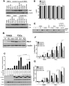
Figure 6. Doxorubicin synergizes with salinomycin to kill LMW-E-expressing tumor cells
(A) Cells were seeded on monolayer for 24 hours and treated with salinomycin for 4 days with the drug-containing media replaced every 48 hours. After 3 days of recovery in drug-free medium, the cells were trypsinized and stained for PE-CD24 and APC-CD44 antibodies and analyzed by FACS analysis. (Error bars = +/- SEM). (B) Cells were seeded on ultra-low attachment plates on day 0 and then treated with salinomycin on days 1 and 3. On day 7, MTT solution was added to the mammosphere cultures and incubated at 37°C for 1 hour. Number of mammospheres were quantified using the automated GelCount program. (Error bars = +/- SEM). (C) Cells were seeded on monolayer for 24 hours and treated with doxorubicin and analyzed by FACS as in (A). (Error bars = +/- SEM). (D) An isobologram illustrates the effects of the combined drugs calculated from the Calcusyn software. The combination indices calculated that lay above the line of additivity represent antagonistic, on the line are additive and underneath the line are synergistic effect. (E) The results obtained from A were subjected to calculation using the Calcusyn software. The isobolograms shown are representative from 3 independent experiments. (F) The combination indices were averaged from 3 independent experiments and the statistical analysis used was unpaired student's t-test (error bars = +/- SEM).

Similar articles

Cited by

References

    1. Koff A, Giordano A, Desai D, Yamashita K, Harper JW, Elledge S, et al. Formation and activation of a cyclin E-cdk2 complex during the G1 phase of the human cell cycle. Science. 1992;257:1689–94. - PubMed
    1. Draetta GF. Mammalian G1 cyclins. Current opinion in cell biology. 1994;6:842–6. - PubMed
    1. Bresnahan WA, Boldogh I, Ma T, Albrecht T, Thompson EA. Cyclin E/Cdk2 activity is controlled by different mechanisms in the G0 and G1 phases of the cell cycle. Cell growth & differentiation : the molecular biology journal of the American Association for Cancer Research. 1996;7:1283–90. - PubMed
    1. Keyomarsi K, Herliczek TW. The role of cyclin E in cell proliferation, development and cancer. Progress in cell cycle research. 1997;3:171–91. - PubMed
    1. Bito T, Ueda M, Ito A, Ichihashi M. Less expression of cyclin E in cutaneous squamous cell carcinomas than in benign and premalignant keratinocytic lesions. Journal of cutaneous pathology. 1997;24:305–8. - PubMed

Publication types

MeSH terms

  NODES
Association 1
twitter 2