. 2013 Oct 22;110(43):17516-21.
doi: 10.1073/pnas.1312374110. Epub 2013 Oct 7.

Pooled RNAi screen identifies ubiquitin ligase Itch as crucial for influenza A virus release from the endosome during virus entry

Affiliations

Pooled RNAi screen identifies ubiquitin ligase Itch as crucial for influenza A virus release from the endosome during virus entry

Wen-Chi Su et al. Proc Natl Acad Sci U S A. .

Abstract

Influenza viruses, like other viruses, rely on host factors to support their life cycle as viral proteins usually "hijack," or collaborate with, cellular proteins to execute their functions. Identification and understanding of these factors can increase the knowledge of molecular mechanisms manipulated by the viruses and facilitate development of antiviral drugs. To this end, we developed a unique genome-wide pooled shRNA screen to search for cellular factors important for influenza A virus (IAV) replication. We identified an E3 ubiquitin ligase, Itch, as an essential factor for an early step in the viral life cycle. In Itch knockdown cells, the incorporation of viral ribonucleoprotein complex into endosomes was normal, but its subsequent release from endosomes and transport to the nucleus was retarded. In addition, upon virus infection, Itch was phosphorylated and recruited to the endosomes, where virus particles were located. Furthermore, Itch interacted with viral M1 protein and ubiquitinated M1 protein. Collectively, our findings unravel a critical role of Itch in mediating IAV release from the endosome and offer insights into the mechanism for IAV uncoating during virus entry. These findings also highlight the feasibility of pooled RNAi screening for exploring the cellular cofactors of lytic viruses.


Conflict of interest statement

The authors declare no conflict of interest.

Figures

Fig. 1.
Fig. 1.
Genome-wide RNAi screen identifies host genes required for IAV replication. (A) Schematic presentation of pooled RNAi screen. (B) Outline of the screening procedures. Numbers of genes associated with the process are shown on the right side. HCS, high-content screening.
Fig. 2.
Fig. 2.
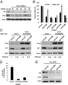
Silencing Itch reduces IAV replication. (A and B) The shRNA-transduced A549 cells were infected with WSN at an MOI of 0.5. At 6 h p.i., the cells were harvested. (A) Immunoblotting against anti-NP and anti-Itch antibodies. Actin was used as loading control. (B) Cellular RNA was extracted and measured by quantitative RT-PCR. The levels of viral RNA and Itch mRNA were normalized by GAPDH mRNA. Values represent the mean ± SD of three independent experiments. (C) A549 cells were transduced with the lentiviruses carrying the Itch wobble mutants to establish stable cell lines and then transduced with shLacZ (as control) or Itch shRNA clone 2 or clone 3 virus as indicated. After selection with puromycin, the shRNA-transduced naïve or stable cells were reseeded and infected with WSN at an MOI of 0.5. Cells were harvested at 6 h p.i. and subjected to Western blot analysis with the indicated antibodies. The band intensities of NP and actin were quantified, and the relative NP/actin ratios are shown below the blots. (D) The shRNA-transduced A549 cells were infected with WSN at an MOI of 0.01. The supernatants were collected at 24 h p.i. and used for determining viral titer by plaque-forming unit assay in MDCK cells. (E) The experiment was performed the same way as in A except that H3N2 virus was used in place of WSN virus.
Fig. 3.
Fig. 3.
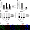
IAV is confined in endosomes in Itch KD cells. (A) The shRNA-transduced A549 cells were infected with WSN strain (MOI 5) for 20 min. The cytoplasmic and nuclear viral RNA was extracted and used for quantitative RT-PCR. Each vRNA level was compared with that of the control, shLacZ. Values represent the mean ± SD of three independent experiments. (B) The shRNA-transduced A549 cells were infected with WSN strain (MOI 5) for 30 min and fixed for immunofluorescence staining with anti-NP antibody and DAPI. Values are shown as numbers of cells displaying nuclear localization of NP among 100 NP-staining cells. Values represent the mean ± SD of three independent experiments. (C) The shRNA-transduced A549 cells and BafA1 (40 nM)-treated cells were infected with WSN (MOI 5) for 10 min or 60 min and then processed for subcellular fractionation. The fractions from different compartments were used for RNA isolation and determination of their vRNA levels by quantitative RT-PCR. The percentage of vRNA was the ratio of the vRNA from each compartment to the vRNA from whole cells. Values represent the mean of two independent experiments. (D) The shRNA-transduced A549 cells were infected with WSN (MOI 5) for 60 min, permeabilized with 0.05% saponin for 2 min, and then fixed for immunofluorescence stain with anti-M1 (green) antibody, anti-Rab7 (red) antibody, and DAPI (blue).The white arrowheads indicate the nuclear distribution of M1.
Fig. 4.
Fig. 4.
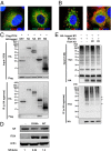
Itch localizes in endosomes upon IAV infection. (A and B) Flag-tagged Itch-expressing A549 cells were infected or uninfected with WSN at an MOI of 50 for 30 min. After virus infection, the cells were processed without (A) or with (B) 0.05% saponin treatment for 2 min, then fixed and stained with anti-Flag antibody (green) and DAPI (blue).
Fig. 5.
Fig. 5.
Itch interacts with M1 and mediates ubiquitination of M1. (A and B) Flag-tagged Itch-expressing A549 cells were infected with WSN at an MOI of 50 for 30 min then treated with 0.05% saponin and processed for immunofluorescence staining with the following antibodies: (A) anti-Flag antibody (green), anti-EEA1 or anti-CD63 antibody (red), and DAPI (blue) or (B) anti-Flag antibody (green), anti-HA or anti-M1 antibody (red), and DAPI (blue). (C) HEK293T cells were cotransfected with Flag-tagged Itch and the indicated HA-tagged plasmids. The cell lysates were prepared and used for immunoprecipitation with HA agarose. The cell lysates or immunoprecipitates were resolved on SDS/PAGE and immunoblotted with anti-HA or anti-Flag antibody. (D) Naïve, WT Itch-overexpressing and C830A Itch mutant-overexpressing A549 cells were infected with WSN at an MOI of 0.5. At 6 h p.i. the cells were harvested for Western blot analysis against anti-NP and anti-Itch antibodies. The relative NP/actin ratios are shown below the blots. (E) The shRNA-transduced HEK293T cells were cotransfected with HA-tagged M1 and Myc-tagged ubiquitin. The lysates were prepared and used for immunoprecipitation with HA agarose. The cell lysates or immunoprecipitates were resolved on SDS/PAGE and immunoblotted with anti-Myc or anti-HA antibody. shLacZ- transduced HEK293T cells were used as control cells. The arrows indicated the mono- and oligo-ubiquitinated M1 species.
Fig. 6.
Fig. 6.
Itch is phosphorylated in the early stage of the IAV life cycle. (A) A549 cells were incubated with WSN (MOI 5) at 4 °C for 1 h and refreshed with warm medium at time 0. The cell extracts were harvested at the indicated time points and analyzed by Western blotting with anti–phospho-Itch or anti-Itch antibody. Actin was used as loading control. (B and C) A549 cells were treated with the inhibitors for 1 h and then infected with WSN (MOI 5) at 4 °C for 1 h in the presence of inhibitors. After virus infection, the cells were refreshed with warm medium and harvested at 30 min or 60 min p.i. The cell extracts were analyzed by Western blotting with anti–phospho-Itch or anti-Itch antibody. The band intensities of P-Itch and actin were quantified, and the relative P-Itch/actin ratios are shown below the blots. The concentrations of inhibitors are as follows: wortmannin, 1 μM; LY294002, 50 μM; BafA1, 40 nM; and NH4Cl, 50 mM.
Fig. 7.
Fig. 7.
The proposed role of Itch in IAV entry. During endocytosis of IAV, Itch becomes phosphorylated and resides in late endosomes. Itch probably mediates IAV release from late endosomes by ubiquitinating M1 to facilitate dissociation of viral RNA from M1 and subsequent transport of vRNP to the nuclei.

Similar articles

Cited by

References

    1. Bouvier NM, Palese P. The biology of influenza viruses. Vaccine. 2008;26(Suppl 4):D49–D53. - PMC - PubMed
    1. Lakadamyali M, Rust MJ, Zhuang X (2004) Endocytosis of influenza viruses. Microbes Infection 6(10):929–936. - PMC - PubMed
    1. Brass AL, et al. The IFITM proteins mediate cellular resistance to influenza A H1N1 virus, West Nile virus, and dengue virus. Cell. 2009;139(7):1243–1254. - PMC - PubMed
    1. Hao L, et al. Drosophila RNAi screen identifies host genes important for influenza virus replication. Nature. 2008;454(7206):890–893. - PMC - PubMed
    1. König R, et al. Human host factors required for influenza virus replication. Nature. 2010;463(7282):813–817. - PMC - PubMed

MeSH terms

  NODES
INTERN 1
twitter 2