Comparison of the condyle-fossa relationship between skeletal class III malocclusion patients with and without asymmetry: a retrospective three-dimensional cone-beam computed tomograpy study
- PMID: 24228235
- PMCID: PMC3822060
- DOI: 10.4041/kjod.2013.43.5.209
Comparison of the condyle-fossa relationship between skeletal class III malocclusion patients with and without asymmetry: a retrospective three-dimensional cone-beam computed tomograpy study
Abstract
Objective: This study investigated whether temporomandibular joint (TMJ) condyle-fossa relationships are bilaterally symmetric in class III malocclusion patients with and without asymmetry and compared to those with normal occlusion. The hypothesis was a difference in condyle-fossa relationships exists in asymmetric patients.
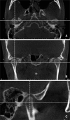
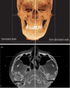
Methods: Group 1 comprised 40 Korean normal occlusion subjects. Groups 2 and 3 comprised patients diagnosed with skeletal class III malocclusion, who were grouped according to the presence of mandibular asymmetry: Group 2 included symmetric mandibles, while group 3 included asymmetric mandibles. Pretreatment three-dimensional cone-beam computed tomography (3D CBCT) images were obtained. Right- and left-sided TMJ spaces in groups 1 and 2 or deviated and non-deviated sides in group 3 were evaluated, and the axial condylar angle was compared.
Results: The TMJ spaces demonstrated no significant bilateral differences in any group. Only group 3 had slightly narrower superior spaces (p < 0.001). The axial condylar angles between group 1 and 2 were not significant. However, group 3 showed a statistically significant bilateral difference (p < 0.001); toward the deviated side, the axial condylar angle was steeper.
Conclusions: Even in the asymmetric group, the TMJ spaces were similar between deviated and non-deviated sides, indicating a bilateral condyle-fossa relationship in patients with asymmetry that may be as symmetrical as that in patients with symmetry. However, the axial condylar angle had bilateral differences only in asymmetric groups. The mean TMJ space value and the bilateral difference may be used for evaluating condyle-fossa relationships with CBCT.
Keywords: Class III diagnosis; Condyle-fossa relationship; Facial asymmetry; TMJ; Three-dimensional cone-beam computed tomography.
Conflict of interest statement
The authors report no commercial, proprietary, or financial interest in the products or companies described in this article.
Figures


Similar articles
-
[Cone-beam CT evaluation of temporomandibular joint in skeletal class Ⅱ female adolescents with different vertical patterns].Beijing Da Xue Xue Bao Yi Xue Ban. 2020 Dec 29;53(1):109-119. doi: 10.19723/j.issn.1671-167X.2021.01.017. Beijing Da Xue Xue Bao Yi Xue Ban. 2020. PMID: 33550344 Free PMC article. Chinese.
-
Three-dimensional analysis of the temporomandibular joint and fossa-condyle relationship.Orthodontics (Chic.). 2011 Fall;12(3):210-21. Orthodontics (Chic.). 2011. PMID: 22022692
-
Comprehensive three-dimensional positional and morphological assessment of the temporomandibular joint in skeletal Class II patients with mandibular retrognathism in different vertical skeletal patterns.BMC Oral Health. 2022 Apr 28;22(1):149. doi: 10.1186/s12903-022-02174-6. BMC Oral Health. 2022. PMID: 35484618 Free PMC article.
-
Three-dimensional characteristics of temporomandibular joint morphology and condylar movement in patients with mandibular asymmetry.Prog Orthod. 2022 Dec 29;23(1):50. doi: 10.1186/s40510-022-00445-0. Prog Orthod. 2022. PMID: 36577877 Free PMC article.
-
[Three-dimensional assessment and study on temporomandibular joint and the maxillary characteristics of skeletal Class Ⅲ mandibular deviation patients].Shanghai Kou Qiang Yi Xue. 2023 Feb;32(1):91-96. Shanghai Kou Qiang Yi Xue. 2023. PMID: 36973851 Chinese.
Cited by
-
Comparison of temporomandibular joints in relation to ages and vertical facial types in skeletal class II female patients: a multiple-cross-sectional study.BMC Oral Health. 2024 Apr 17;24(1):467. doi: 10.1186/s12903-024-04219-4. BMC Oral Health. 2024. PMID: 38632555 Free PMC article.
-
Facial asymmetry of the hard and soft tissues in skeletal Class I, II, and III patients.Sci Rep. 2024 Feb 29;14(1):4966. doi: 10.1038/s41598-024-55107-4. Sci Rep. 2024. PMID: 38424179 Free PMC article.
-
Assessment of the Morphology and Degenerative Changes in the Temporomandibular Joint Using CBCT according to the Orthodontic Approach: A Scoping Review.Biomed Res Int. 2022 Feb 1;2022:6863014. doi: 10.1155/2022/6863014. eCollection 2022. Biomed Res Int. 2022. PMID: 35155678 Free PMC article. Review.
-
Auto exposure control (AEC) modality in CBCT devices: Revisiting the reported parameters in current orthodontic literature.J Oral Biol Craniofac Res. 2015 Sep-Dec;5(3):236-7. doi: 10.1016/j.jobcr.2015.06.006. Epub 2015 Aug 1. J Oral Biol Craniofac Res. 2015. PMID: 26605149 Free PMC article. No abstract available.
-
Comparison of maxillomandibular asymmetries in adult patients presenting different sagittal jaw relationships.Dental Press J Orthod. 2019 Sep 5;24(4):54-62. doi: 10.1590/2177-6709.24.4.054-062.oar. Dental Press J Orthod. 2019. PMID: 31508707 Free PMC article.
References
-
- Katsavrias EG, Halazonetis DJ. Condyle and fossa shape in Class II and Class III skeletal patterns: a morphometric tomographic study. Am J Orthod Dentofacial Orthop. 2005;128:337–346. - PubMed
-
- Ahn SJ, Lee SP, Nahm DS. Relationship between temporomandibular joint internal derangement and facial asymmetry in women. Am J Orthod Dentofacial Orthop. 2005;128:583–591. - PubMed
-
- Byun ES, Ahn SJ, Kim TW. Relationship between internal derangement of the temporomandibular joint and dentofacial morphology in women with anterior open bite. Am J Orthod Dentofacial Orthop. 2005;128:87–95. - PubMed
-
- Vitral RW, da Silva Campos MJ, Rodrigues AF, Fraga MR. Temporomandibular joint and normal occlusion: Is there anything singular about it? A computed tomographic evaluation. Am J Orthod Dentofacial Orthop. 2011;140:18–24. - PubMed
-
- Rodrigues AF, Fraga MR, Vitral RW. Computed tomography evaluation of the temporomandibular joint in Class I malocclusion patients: condylar symmetry and condyle-fossa relationship. Am J Orthod Dentofacial Orthop. 2009;136:192–198. - PubMed
LinkOut - more resources
Full Text Sources
Other Literature Sources
