Tau causes synapse loss without disrupting calcium homeostasis in the rTg4510 model of tauopathy
- PMID: 24278327
- PMCID: PMC3835324
- DOI: 10.1371/journal.pone.0080834
Tau causes synapse loss without disrupting calcium homeostasis in the rTg4510 model of tauopathy
Abstract
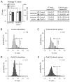
Neurofibrillary tangles (NFTs) of tau are one of the defining hallmarks of Alzheimer's disease (AD), and are closely associated with neuronal degeneration. Although it has been suggested that calcium dysregulation is important to AD pathogenesis, few studies have probed the link between calcium homeostasis, synapse loss and pathological changes in tau. Here we test the hypothesis that pathological changes in tau are associated with changes in calcium by utilizing in vivo calcium imaging in adult rTg4510 mice that exhibit severe tau pathology due to over-expression of human mutant P301L tau. We observe prominent dendritic spine loss without disruptions in calcium homeostasis, indicating that tangles do not disrupt this fundamental feature of neuronal health, and that tau likely induces spine loss in a calcium-independent manner.
Conflict of interest statement
Figures


Similar articles
-
Synaptic alterations in the rTg4510 mouse model of tauopathy.J Comp Neurol. 2013 Apr 15;521(6):1334-53. doi: 10.1002/cne.23234. J Comp Neurol. 2013. PMID: 23047530 Free PMC article.
-
Early depletion of CA1 neurons and late neurodegeneration in a mouse tauopathy model.Brain Res. 2017 Jun 15;1665:22-35. doi: 10.1016/j.brainres.2017.04.002. Epub 2017 Apr 11. Brain Res. 2017. PMID: 28411086
-
Sex Impact on Tau-Aggregation and Postsynaptic Protein Levels in the P301L Mouse Model of Tauopathy.J Alzheimers Dis. 2017;56(4):1279-1292. doi: 10.3233/JAD-161087. J Alzheimers Dis. 2017. PMID: 28157099
-
Tauopathies and tau oligomers.J Alzheimers Dis. 2013;37(3):565-8. doi: 10.3233/JAD-130653. J Alzheimers Dis. 2013. PMID: 23948895 Review.
-
Synaptic Localisation of Tau.Adv Exp Med Biol. 2019;1184:105-112. doi: 10.1007/978-981-32-9358-8_9. Adv Exp Med Biol. 2019. PMID: 32096032 Review.
Cited by
-
Synapses in neurodegenerative diseases.BMB Rep. 2017 May;50(5):237-246. doi: 10.5483/bmbrep.2017.50.5.038. BMB Rep. 2017. PMID: 28270301 Free PMC article. Review.
-
Hyperphosphorylated tau causes reduced hippocampal CA1 excitability by relocating the axon initial segment.Acta Neuropathol. 2017 May;133(5):717-730. doi: 10.1007/s00401-017-1674-1. Epub 2017 Jan 16. Acta Neuropathol. 2017. PMID: 28091722 Free PMC article.
-
Experimental evidence for the age dependence of tau protein spread in the brain.Sci Adv. 2019 Jun 26;5(6):eaaw6404. doi: 10.1126/sciadv.aaw6404. eCollection 2019 Jun. Sci Adv. 2019. PMID: 31249873 Free PMC article.
-
The intersection of amyloid beta and tau at synapses in Alzheimer's disease.Neuron. 2014 May 21;82(4):756-71. doi: 10.1016/j.neuron.2014.05.004. Neuron. 2014. PMID: 24853936 Free PMC article. Review.
-
Cholinergic activity is essential for maintaining the anterograde transport of Choline Acetyltransferase in Drosophila.Sci Rep. 2018 May 23;8(1):8028. doi: 10.1038/s41598-018-26176-z. Sci Rep. 2018. PMID: 29795337 Free PMC article.
References
Publication types
MeSH terms
Substances
Grants and funding
- P50AG05134/AG/NIA NIH HHS/United States
- AG026249/AG/NIA NIH HHS/United States
- P30 AG062421/AG/NIA NIH HHS/United States
- S10 RR025645/RR/NCRR NIH HHS/United States
- R01 AG008487/AG/NIA NIH HHS/United States
- T32 AG000277/AG/NIA NIH HHS/United States
- R01 EB000768/EB/NIBIB NIH HHS/United States
- P50 AG005134/AG/NIA NIH HHS/United States
- T32AG000277/AG/NIA NIH HHS/United States
- R00 AG033670/AG/NIA NIH HHS/United States
- R01 AG026249/AG/NIA NIH HHS/United States
- R00AG33670/AG/NIA NIH HHS/United States
- AG08487/AG/NIA NIH HHS/United States
LinkOut - more resources
Full Text Sources
Other Literature Sources
Molecular Biology Databases

