. 2014 Jan 16;156(1-2):45-68.
doi: 10.1016/j.cell.2013.12.019.

Reversing DNA methylation: mechanisms, genomics, and biological functions

Affiliations

Reversing DNA methylation: mechanisms, genomics, and biological functions

Hao Wu et al. Cell. .

Abstract

Methylation of cytosines in the mammalian genome represents a key epigenetic modification and is dynamically regulated during development. Compelling evidence now suggests that dynamic regulation of DNA methylation is mainly achieved through a cyclic enzymatic cascade comprised of cytosine methylation, iterative oxidation of methyl group by TET dioxygenases, and restoration of unmodified cytosines by either replication-dependent dilution or DNA glycosylase-initiated base excision repair. In this review, we discuss the mechanism and function of DNA demethylation in mammalian genomes, focusing particularly on how developmental modulation of the cytosine-modifying pathway is coupled to active reversal of DNA methylation in diverse biological processes.


Figures

Figure 1
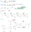
Figure 1. Domain architecture and enzymatic activities of cytosine methylation and demethylation machineries
(A) Schematic diagrams of predicted functional domains in proteins involved in de novo methylation, maintenance methylation, 5mC oxidation, and 5fC/5caC excision. All TET proteins contain a C-terminal catalytic domain that includes a Cys-rich insert and a large double-stranded β-helix (DSBH) domain. TET1 and TET3 proteins also contain a N-terminal CXXC domain. TDG contains a uracil DNA glycosylase (UDG) domain. The number of amino acids of the full-length isoform is indicated, and the numbering corresponds to mouse proteins. ADD, ATRX-DNMT3-DNMT3L; PWWP, proline-tryptophan-tryptophan-proline motif; MTase, DNA methyltransferase domain; BAH, bromo-adjacent homology; CXXC, zinc-finger Cystien-X-X-Cystine; SRA, SET and RING associated; RING, Really interesting new gene; PHD, Plant homeodomain; UBL, ubiquitin-like domain. (B) The step-wise cytosine modification pathway that includes cytosine methylation by DNMTs and iterative oxidation of 5mC by TET proteins. DNMTs use S-adenosylmethionine (SAM) as a methyl donor to catalyze methylation at the 5-position of cytosine, yielding S-adenosylhomocysteine (SAH). As Fe(II)/α-ketoglutarate (α-KG)-dependnet dioxygenases, TET proteins use a base-flipping mechanism to flip the _target base out of the duplex DNA into the catalytic site. The active site Fe(II) is bound by conserved His-His-Asp residues in TETs and coordinates water and α-KG. TET enzymes use molecular oxygen as a substrate to catalyze oxidative decarboxylation of αKG, yielding CO2, enzyme-bound succinate, and a reactive high-valent Fe(IV)-oxo intermediate. The enzyme-bound Fe(IV)-oxo intermediate reacts with 5mC/5hmC/5fC to generate 5hmC/5fC/5caC, with a net oxidative transfer of the single oxygen atom to the substrate, resulting in regeneration of the Fe(II) species. (C) Base excision repair of 5mC (in plants) or 5fC/5caC (in mammals) to complete DNA demethylation. In plants, the DME/ROS1 family of bifunctional DNA glycosylases is capable of catalyzing the release of 5mC base from DNA by cleavage of the N-glycosidic bond, generating an abasic [apurinic and apyrimidinic (AP)] site. The DNA backbone is then nicked by AP lyase activity of DME/ROS1 enzymes. The 3′ sugar group is then cleaved by an AP endonuclease and the resulting single-nucleotide gap is filled in with an unmodified C by DNA polymerase and ligase activities. In mammals, oxidized 5mC bases (5fC/5caC), could be excised by TDG. TDG is a monofunctional DNA glycosylase, so other proteins are required to provide the AP lyase activity to remove the sugar ring to generate a single-nucleotide gap.
Figure 2
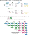
Figure 2. Mechanisms of passive and active reversal of CpG DNA methylation
(A) Schematic diagrams of replication-dependent passive loss of 5mC or oxidized 5mC within CpG dyads. Replication-dependent passive dilution (PD) of 5mC occurs in the absence of the DNA methylation maintenance machinery (DNMT1/UHRF1). By contrast, 5mC oxidation derivatives, 5hmC (h) [potentially 5fC (f) and 5caC (ca)], may facilitate passive demethylation as hemi-hydroxymethylated CpGs is an inefficient substrate for DNMT1. This form of active DNA demethylation is termed as active modification (AM) followed by passive dilution (AM-PD). Whether DNMT1 or DNMT3 can efficiently methylate CpG dyads hemi-modified with 5fC/5caC is unclear. (B) Schematic diagrams of replication-independent DNA demethylation within CpG dyads. TET and TDG mediate sequential 5mC oxidation and 5fC/5caC excision. The resulting abasic site is repaired by BER to regenerate unmodified cytosines. All possible intermediate products (a total of 21 distinct cytosine modification states for a single CpG dyad) of the DNMT/TET/TDG/BER enzymatic cascade are depicted. For each state, the left half represents the top strand of CpG dyad, whereas the right half indicates the bottom strand. The arrows represent enzymatic reactions and are color-coded according to that of the corresponding enzyme. One of many potential combinations of intermediate steps for the TET/TDG-mediated active DNA demethylation process is highlighted in gray. Of note, 5fC can be directly excised by TDG to generate abasic site (e.g. f:ca -> ab:ab). For clarity, all f->ab reactions are not depicted.
Figure 3
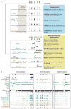
Figure 3. Genome-wide mapping methods for cytosine modifications
(A) Summary of BS-Seq or affinity-enrichment based genome-wide mapping methods of DNA cytosine modifications. In conjunction with various chemical and enzymatic pre-treatment, BS-Seq or affinity-enrichment (antibody or chemical tagging) methods have been developed to map 5mC (BS-Seq and oxBS-Seq; 5mC DIP), 5hmC (oxBS-seq and TAB-Seq; 5hmC DIP, CMS IP, JBP1 IP, GLIB, and hMe-Seal), 5fC (fCAB-Seq; 5fC DIP, fC-Seal, and 5fC pull-down), and 5caC (caCAB-Seq; 5caC DIP) (see Box 2). Notably, oxBS-Seq, fCAB-Seq, and caCAB-Seq require subtracting signals of conventional BS-Seq from those of modified BS-Seq to indirectly determine the position and abundance of oxidized 5mC bases. 5fC and 5caC have not been systematically mapped at base-resolution, as they are relatively rare in the wild-type cells and require unusually high sequencing coverage to confidently determine their position and abundance. (B) Shown are genomic distributions of oxidized 5mC bases at representative loci in mouse ESCs. Genomic distribution of 5hmC/5fC/5caC in control (wt: wild-type; Ctrl kd: control knockdown) and Tdg-depleted (ko: knockout; Tdg kd: Tdg knockdown) mouse ESCs are shown for three representative loci. 5hmC distribution was measured by both affinity-based method [5hmC DIP, CMS, 5hmC-seal, and GLIB] (Pastor et al., 2011; Shen et al., 2013; Song et al., 2013a) and base-resolution method [TAB-Seq, scale: 0–50%] (Yu et al., 2012). Distributions of 5fC and 5caC were determined by antibody [5fC/5caC DIP] (Shen et al., 2013) or chemical tagging [5fC-seal] (Song et al., 2013a) methods. DNA methylation levels were estimated by 5mC DIP (Shen et al., 2013) or conventional BS-Seq (5mC+5hmC, scale: 0–100%) in wild-type mouse ESCs (Stadler et al., 2011). Based on local methylation levels, the genome can be categorized into FMRs (Fully methylated regions, 50–100% methylation), LMRs (low methylated regions, 10–50%), and UMRs (unmethylated regions, <10%) (Stadler et al., 2011). Also shown are other genomic datasets in wild-type mouse ESCs, including TET1 ChIP-seq (Wu et al., 2011b), RNA-seq (Ficz et al., 2011), major histone modifications (Mikkelsen et al., 2007), DNase I hypersensitivity site (modENCODE project) and binding sites of pluripotency TFs (Whyte et al., 2012). A region located upstream of Zfp281 gene is highlighted in gray, where Tdg-deficiency induced ectopic 5caC is overlapped with chromatin features of active enhancer (DNase I+/H3K4me1+/H3K4me3) and binding sites of pluripotency factors (Oct4/Nanog/Sox2).
Figure 4
Figure 4. DNA methylation dynamics during mouse preimplantation development
(A) Dynamic changes in cytosine modifications and relevant enzymes during preimplantation development. Immediately after fertilization, paternal 5mC is rapidly oxidized to 5hmC/5fC/5caC by TET3 proteins. In early pre-implantation embryos, oocyte-derived DNMT1o is largely excluded from nucleus (dash line) and consequently maintenance methylation is inefficient. Oxidized 5mC bases in the paternal genome and 5mC in the maternal genome are therefore passively diluted. The global 5mC level reaches the lowest point around blastocyst stage (E3.5). After implantation, DNA methylation pattern is reestablished by DNMT3A/3B in inner cell mass (ICM) cells, but not in trophectoderm (TE) cells. The genome of ground-state ESC (2i-condition) is hypomethylated (5mC: 1% of all C) and is more similar to the methylome of pre-implantation ICM cells, whereas primed ESCs (serum) possess a methylome (5mC: 4% of all C) that recapitulates overall methylation pattern in epiblast cells. (B) Developmental stage-specific usage of the cyclic cytosine modifying enzymatic cascade during early embryonic development. Upon fertilization, oocyte-derived TET3 proteins specifically localize to paternal nucleus and convert sperm-derived 5mC to 5hmC/5fC/5caC. DNA replication drives passive loss of 5hmC/5fC/5caC in the paternal genome and 5mC in the maternal genome (left panel). In primed ESCs and cells during ICM-to-epiblast transition in vivo, all enzymes of the cytosine modifying cascade are expressed at relatively high levels. Dnmt3A/3B/3L and DNMT1/UHRF1 establish and maintain 5mC patterns. At some genomic regions (e.g. active enhancers and bivalent promoters) in these cells, TET1/2 oxidize 5mC to 5hmC/5fC/5caC, and TDG/BER excises 5fC/5caC to regenerate unmodified cytosine.
Figure 5
Figure 5. Global reprogramming of DNA methylation patterns during early PGC development
(A) Dynamic changes in cytosine modifications and expression levels of relevant enzymes during early stage of mouse PGC development. Global erasure of DNA methylation in developing PGCs goes through two phases. In the first phase (E7.25 to E9.0), both de novo and maintenance methylation machineries are functionally impaired due to the repression of DNMT3A/3B/3L and UHRF1. There is also evidence that remaining UHRF1 is excluded from nucleus (dash line). Thus, loss of bulk genomic DNA methylation (solid line, inherited from epiblast cell stage) takes place in a replication-dependent manner in phase 1. However, a portion of the genome (dash line) including ICRs and germline-specific genes are partially or fully protected from the first wave of passive demethylation. In the second phase (E9.5 to E12.0), TET1 (possibly TET2) are up-regulated and oxidize remaining 5mC to 5hmC. (B) Developmental stage-specific usage of the cytosine modifying enzymatic cascade during early stage of PGC development. In phase 1 of PGC demethylation, bulk of the genome undergoes global demethylation in the absence of de novo and maintenance DNA methylation. In phase 2, TET1/2 proteins oxidize remaining 5mC at late demethylating loci. Replication-dependent loss of 5hmC completes the global demethylation process. 5fC/5caC is not specifically enriched in developing PGCs as compared to surrounding somatic cells, so they may be actively excised by TDG/BER pathway that is present in PGCs.

Similar articles

Cited by

References

    1. Barreto G, Schafer A, Marhold J, Stach D, Swaminathan SK, Handa V, Doderlein G, Maltry N, Wu W, Lyko F, et al. Gadd45a promotes epigenetic gene activation by repair-mediated DNA demethylation. Nature. 2007;445:671–675. - PubMed
    1. Baylin SB, Jones PA. A decade of exploring the cancer epigenome - biological and translational implications. Nat Rev Cancer. 2011;11:726–734. - PMC - PubMed
    1. Bennett MT, Rodgers MT, Hebert AS, Ruslander LE, Eisele L, Drohat AC. Specificity of human thymine DNA glycosylase depends on N-glycosidic bond stability. J Am Chem Soc. 2006;128:12510–12519. - PMC - PubMed
    1. Bernstein BE, Mikkelsen TS, Xie X, Kamal M, Huebert DJ, Cuff J, Fry B, Meissner A, Wernig M, Plath K, et al. A bivalent chromatin structure marks key developmental genes in embryonic stem cells. Cell. 2006;125:315–326. - PubMed
    1. Bhutani N, Brady JJ, Damian M, Sacco A, Corbel SY, Blau HM. Reprogramming towards pluripotency requires AID-dependent DNA demethylation. Nature. 2010;463:1042–1047. - PMC - PubMed

Publication types

Substances

  NODES
Note 1
Project 1
twitter 2