Epidermal Th22 and Tc17 cells form a localized disease memory in clinically healed psoriasis
- PMID: 24610014
- PMCID: PMC3962894
- DOI: 10.4049/jimmunol.1302313
Epidermal Th22 and Tc17 cells form a localized disease memory in clinically healed psoriasis
Abstract
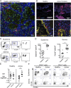
Psoriasis is a common and chronic inflammatory skin disease in which T cells play a key role. Effective treatment heals the skin without scarring, but typically psoriasis recurs in previously affected areas. A pathogenic memory within the skin has been proposed, but the nature of such site-specific disease memory is unknown. Tissue-resident memory T (TRM) cells have been ascribed a role in immunity after resolved viral skin infections. Because of their localization in the epidermal compartment of the skin, TRM may contribute to tissue pathology during psoriasis. In this study, we investigated whether resolved psoriasis lesions contain TRM cells with the ability to maintain and potentially drive recurrent disease. Three common and effective therapies, narrowband-UVB treatment and long-term biologic treatment systemically inhibiting TNF-α or IL-12/23 signaling were studied. Epidermal T cells were highly activated in psoriasis and a high proportion of CD8 T cells expressed TRM markers. In resolved psoriasis, a population of cutaneous lymphocyte-associated Ag, CCR6, CD103, and IL-23R expressing epidermal CD8 T cells was highly enriched. Epidermal CD8 T cells expressing the TRM marker CD103 responded to ex vivo stimulation with IL-17A production and epidermal CD4 T cells responded with IL-22 production after as long as 6 y of TNF-α inhibition. Our data suggest that epidermal TRM cells are retained in resolved psoriasis and that these cells are capable of producing cytokines with a critical role in psoriasis pathogenesis. We provide a potential mechanism for a site-specific T cell-driven disease memory in psoriasis.
Figures


Similar articles
-
Significance of IL-17A-producing CD8+CD103+ skin resident memory T cells in psoriasis lesion and their possible relationship to clinical course.J Dermatol Sci. 2019 Jul;95(1):21-27. doi: 10.1016/j.jdermsci.2019.06.002. Epub 2019 Jun 20. J Dermatol Sci. 2019. PMID: 31300254
-
Tissue-Resident Memory CD8+ T Cells From Skin Differentiate Psoriatic Arthritis From Psoriasis.Arthritis Rheumatol. 2021 Jul;73(7):1220-1232. doi: 10.1002/art.41652. Epub 2021 May 25. Arthritis Rheumatol. 2021. PMID: 33452865 Free PMC article.
-
Immunological Memory in Imiquimod-Induced Murine Model of Psoriasiform Dermatitis.Int J Mol Sci. 2020 Sep 30;21(19):7228. doi: 10.3390/ijms21197228. Int J Mol Sci. 2020. PMID: 33007963 Free PMC article.
-
Immunological Memory of Psoriatic Lesions.Int J Mol Sci. 2020 Jan 17;21(2):625. doi: 10.3390/ijms21020625. Int J Mol Sci. 2020. PMID: 31963581 Free PMC article. Review.
-
Tissue-resident memory T cells in the skin.Inflamm Res. 2020 Mar;69(3):245-254. doi: 10.1007/s00011-020-01320-6. Epub 2020 Jan 27. Inflamm Res. 2020. PMID: 31989191 Review.
Cited by
-
Dihydroartemisinin ameliorates psoriatic skin inflammation and its relapse by diminishing CD8+ T-cell memory in wild-type and humanized mice.Theranostics. 2020 Aug 21;10(23):10466-10482. doi: 10.7150/thno.45211. eCollection 2020. Theranostics. 2020. PMID: 32929360 Free PMC article.
-
Tattooing in Psoriasis: A Questionnaire-Based Analysis of 150 Patients.Clin Cosmet Investig Dermatol. 2022 Apr 6;15:587-593. doi: 10.2147/CCID.S348165. eCollection 2022. Clin Cosmet Investig Dermatol. 2022. PMID: 35418768 Free PMC article.
-
Tissue Resident CD8 Memory T Cell Responses in Cancer and Autoimmunity.Front Immunol. 2018 Nov 29;9:2810. doi: 10.3389/fimmu.2018.02810. eCollection 2018. Front Immunol. 2018. PMID: 30555481 Free PMC article. Review.
-
Tissue-resident memory T cells and their biological characteristics in the recurrence of inflammatory skin disorders.Cell Mol Immunol. 2020 Jan;17(1):64-75. doi: 10.1038/s41423-019-0291-4. Epub 2019 Oct 8. Cell Mol Immunol. 2020. PMID: 31595056 Free PMC article. Review.
-
Resolution of plaque-type psoriasis: what is left behind (and reinitiates the disease).Semin Immunopathol. 2019 Nov;41(6):633-644. doi: 10.1007/s00281-019-00766-z. Epub 2019 Oct 31. Semin Immunopathol. 2019. PMID: 31673756 Free PMC article. Review.
References
-
- Conrad C., Boyman O., Tonel G., Tun-Kyi A., Laggner U., de Fougerolles A., Kotelianski V., Gardner H., Nestle F. O. 2007. α1β1 integrin is crucial for accumulation of epidermal T cells and the development of psoriasis. Nat. Med. 13: 836–842. - PubMed
-
- Lowes M. A., Kikuchi T., Fuentes-Duculan J., Cardinale I., Zaba L. C., Haider A. S., Bowman E. P., Krueger J. G., Zaba L. C., Zaba L. C., et al. 2008. Psoriasis vulgaris lesions contain discrete populations of Th1 and Th17 T cells. J. Invest. Dermatol. 128: 1207–1211. - PubMed
-
- Leonardi C. L., Kimball A. B., Papp K. A., Yeilding N., Guzzo C., Wang Y., Li S., Dooley L. T., Gordon K. B., PHOENIX 1 study investigators 2008. Efficacy and safety of ustekinumab, a human interleukin-12/23 monoclonal antibody, in patients with psoriasis: 76-week results from a randomised, double-blind, placebo-controlled trial (PHOENIX 1). Lancet 371: 1665–1674. - PubMed
Publication types
MeSH terms
Substances
LinkOut - more resources
Full Text Sources
Other Literature Sources
Medical
Research Materials

