Agreement in identification of glaucomatous progression between the optic disc photography and Heidelberg retina tomography in young glaucomatous patients
- PMID: 24967194
- PMCID: PMC4067662
- DOI: 10.3980/j.issn.2222-3959.2014.03.16
Agreement in identification of glaucomatous progression between the optic disc photography and Heidelberg retina tomography in young glaucomatous patients
Abstract
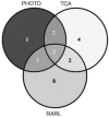
Aim: To evaluate concordance between the clinical assessment of glaucomatous progression of the optic disc photography and progression identified by Heidelberg Retina Tomograph (HRT) in patients with suspected primary juvenile open angle glaucoma (JOAG).
Methods: Optic disc photographs and corresponding HRT II series were reviewed. Optic disc changes between first and final photographs were noted as well as progression identified by HRT topographic change analysis (TCA) and rim area regression line (RARL) Agreement between progression indentified by photography and HRT methods was assessed. Progression, determined from optic disc photographs by consensus assessment was used as the reference standard.
Results: A total of 31 patients (59 eyes) with suspected JOAG were studied. Agreement for progression/no progression between TCA and photography was obtained in 4 progressing eyes and 38 stable eyes (71.19%, k=0.11). Agreement for progression/no progression between RARL and photography was detected in 5 progressing eyes and in 34 stable eyes (66.10%, k=0.15). The number of HRT per patient was statistically higher in the progressing group (P=0.034).
Conclusion: Agreement for detection of longitudinal changes between photography and HRT analysis was poor. One way to improve the chance of discovery of the progression could be increasing the number of HRT examinations.
Keywords: Heidelberg Retina Tomography; juvenile glaucoma; optic disc.
Figures


Similar articles
-
Glaucomatous progression in series of stereoscopic photographs and Heidelberg retina tomograph images.Arch Ophthalmol. 2010 May;128(5):560-8. doi: 10.1001/archophthalmol.2010.52. Arch Ophthalmol. 2010. PMID: 20457976 Free PMC article.
-
Clinicians agreement in establishing glaucomatous progression using the Heidelberg retina tomograph.Ophthalmology. 2009 Jan;116(1):14-24. doi: 10.1016/j.ophtha.2008.08.030. Epub 2008 Nov 17. Ophthalmology. 2009. PMID: 19010552 Free PMC article.
-
Glaucoma follow-up by the Heidelberg retina tomograph--new graphical analysis of optic disc topography changes.Graefes Arch Clin Exp Ophthalmol. 2006 Jun;244(6):654-62. doi: 10.1007/s00417-005-0107-3. Epub 2005 Oct 12. Graefes Arch Clin Exp Ophthalmol. 2006. PMID: 16220279
-
[Stereometric parameters of the optic disc. Comparison between a simultaneous non-mydriatic stereoscopic fundus camera (KOWA WX 3D) and the Heidelberg scanning laser ophthalmoscope (HRT IIII)].Ophthalmologe. 2011 Oct;108(10):957-62. doi: 10.1007/s00347-011-2416-8. Ophthalmologe. 2011. PMID: 21904837 German.
-
Determinants of agreement between the confocal scanning laser tomograph and standardized assessment of glaucomatous progression.Ophthalmology. 2010 Oct;117(10):1953-9. doi: 10.1016/j.ophtha.2010.02.002. Epub 2010 Jun 16. Ophthalmology. 2010. PMID: 20557941 Free PMC article.
Cited by
-
Frequency of Agreement Between Structural and Functional Glaucoma Testing: A Longitudinal Study of 3D OCT and Current Clinical Tests.Am J Ophthalmol. 2024 Oct;266:196-205. doi: 10.1016/j.ajo.2024.05.018. Epub 2024 May 27. Am J Ophthalmol. 2024. PMID: 38810864
References
-
- Feiti ME, Krupin T, Tanna AP. Juvenile glaucoma. In: Roz FH, Fraunfelder FW, Fraunfelder FT, editors. Current Ocular therapy. Amsterdam, The Netherlands: Elsevier; 2008. pp. 500–502.
-
- Weinreb RN, Zangwill LM, Jain S, Becerra LM, Dirkes K, Piltz-Seymour JR, Cioffi GA, Trick GL, Coleman AL, Brandt JD, Liebmann JM, Gordon MO, Kass MA. Predicting the onset of glaucoma: the confocal scanning laser ophthalmoscopy ancillary study to the Ocular Hypertension Treatment Study. Ophthalmology. 2010;117(9):1674–1683. - PMC - PubMed
-
- Zangwill LM, Weinreb RN, Beiser JA, Berry CC, Cioffi GA, Coleman AL, Trick G, Liebmann JM, Brandt JD, Piltz-Seymour JR, Dirkes KA, Vega S, Kass MA, Gordon MO. Baseline topographic optic disc measurements are associated with the development of primary open-angle glaucoma: The Confocal Scanning Laser Ophthalmoscopy Ancillary Study to the Ocular Hypertension Treatment Study. Arch Ophthalmol. 2005;123(9):1188–1197. - PubMed
-
- Chauhan BC, Hutchison DM, Artes PH, Caprioli J, Jonas JB, LeBlanc RP, Nicolela MT. Optic disc progression in glaucoma: comparison of confocal scanning laser tomography to optic disc photographs in a prospective study. Invest Ophthalmol Vis Sci. 2009;50(4):1682–1691. - PubMed
LinkOut - more resources
Full Text Sources
